Bacillus anthracis
Kingdom: Bacteria
Phylum: Firmicutes
Class: Bacilli
Order: Bacillales
Family: Bacillaceae
Genus: Bacillus
Species: anthracis
Kingdom: Bacteria
Phylum: Firmicutes
Class: Bacilli
Order: Bacillales
Family: Bacillaceae
Genus: Bacillus
Species: anthracis
The anthrax bacillus, Bacillus anthracis, was the first bacterium shown to be the cause of a disease. In 1877, Robert Koch grew the organism in pure culture, demonstrated its ability to form endospores, and produced experimental anthrax by injecting it into animals.

Figure 1. Robert Koch's original photomicrographs of Bacillus anthracis, the agent of anthrax. Compare the cell morphology and spore position with the Gram stain below (Figure 2). This is Bacillus anthracis. Beware of phony and mislabeled images of B. anthracis on the internet, including some that are posted by otherwise credible websites. Look for large cells with square ends and centrally-located ellipsoid spores when identifying Bacillus anthracis.
Bacillus anthracis is very large, Gram-positive, sporeforming rod, 1 - 1.2µm in width x 3 - 5µm in length. The bacterium can be cultivated in ordinary nutrient medium under aerobic or anaerobic conditions. Genotypically and phenotypically it is very similar to Bacillus cereus, which is found in soil habitats around the world, and to Bacillus thuringiensis, the pathogen for larvae of Lepidoptera. The three species have the same cellular size and morphology and form oval spores located centrally in a nonswollen sporangium.

Figure 2. Bacillus anthracis. Gram stain. 1500X. The cells have characteristic squared ends. The endospores are ellipsoidal shaped and located centrally in the sporangium. The spores are highly refractile to light and resistant to staining.
Bacillus thuringiensis is distinguished from B. cereus or B. anthracis by its pathogenicity for Lepidopteran insects (moths and caterpillars) and by production of an intracellular parasporal crystal in association with spore formation. The bacteria and protein crystals are sold as "Bt" insecticide, which is used for the biological control of certain garden and crop pests.
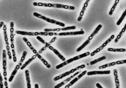
Figure 3. Bacillus thuringiensis. Phase Photomicrograph of vegetative cells, intracellular spores (light) and parasporal crystals (dark). 1000X.
Bacillus cereus is a normal inhabitant of the soil, but it can be regularly isolated from foods such as grains and spices. B. cereus causes two types of food-borne intoxications (as opposed to infections). One type is characterized by nausea and vomiting and abdominal cramps and has an incubation period of 1 to 6 hours. It resembles Staphylococcus aureus food poisoning in its symptoms and incubation period. This is the "short-incubation" or emetic form of the disease. The second type is manifested primarily by abdominal cramps and diarrhea with an incubation period of 8 to 16 hours. Diarrhea may be a small volume or profuse and watery. This type is referred to as the "long-incubation" or diarrheal form of the disease, and it resembles food poisoning caused by Clostridium perfringens. In either type, the illness usually lasts less than 24 hours after onset.
The short-incubation form is caused by a preformed, heat-stable emetic toxin, ETE. The mechanism and site of action of this toxin are unknown, although the small molecule forms ion channels and holes in membranes. The long-incubation form of illness is mediated by the heat-labile diarrheagenic enterotoxin Nheand/or hemolytic enterotoxin HBL, which cause intestinal fluid secretion, probably by several mechanisms, including pore formation and activation of adenylate cyclase enzymes.
The short-incubation form is caused by a preformed, heat-stable emetic toxin, ETE. The mechanism and site of action of this toxin are unknown, although the small molecule forms ion channels and holes in membranes. The long-incubation form of illness is mediated by the heat-labile diarrheagenic enterotoxin Nheand/or hemolytic enterotoxin HBL, which cause intestinal fluid secretion, probably by several mechanisms, including pore formation and activation of adenylate cyclase enzymes.

Figure 4. Bacillus cereus. Gram stain. 450X. Bacilli are large bacteria, so that they are readily observed with the microscope's "high dry objective" ........but you can't detect anything about their spores. This could be a Lactobacillus.
Cultivation
Several nonselective and selective media for the detection and isolation ofBacillus anthracis have been described, as well as a rapid screening test for the bacterium based on the morphology of microcolonies. Table 1 provides the differential characteristics that are used to distinguish Bacillus anthracis from most strains of Bacillus cereus and Bacillus thuringiensis but not necessarily from other saprophytic species of Bacillus. Otherwise, it is not the intent of this article to provide information on the growth of the bacterium in the laboratory.
Table 1. Differential Characteristics of B. anthracis B. cereus and B. thuringiensis
| Characteristic | B. anthracis | B. cereus and B. thuringiensis |
| growth requirement for thiamin | ||
| hemolysis on sheep blood agar | ||
| glutamyl-polypeptide capsule | ||
| lysis by gamma phage | ||
| motility | ||
| growth on chloral hydrate agar | ||
| string-of-pearls test |
The following figures (5, 6, and 7) from the CDC are reliable images ofBacillus anthracis grown as described in the figure legends.

Figure 5. Colonies of Bacillus cereus on the left; colonies of Bacillus anthracis on the right. B. cereus colonies are larger, more mucoid, and this strain exhibits a slight zone of hemolysis on blood agar.
Figure 5. Colonies of Bacillus cereus on the left; colonies of Bacillus anthracis on the right. B. cereus colonies are larger, more mucoid, and this strain exhibits a slight zone of hemolysis on blood agar.

Figure 6. Lysis of Bacillus anthracis by the lytic phage gamma. The plaque (clear area) in the region of confluent growth is where the gamma phage was applied. The plaque results from the phage's ability to lyse the bacterial cells. Since the gamma phage is specific for B. anthracis, and will not lyse B. thuringiensis or B. cereus, we know that this is Bacillus anthracis. The colony type of is similar to Figure 5.

Figure 7. Mucoid colonies of Bacillus anthracis. This culture was probably incubated at an increased CO2 tension (5% CO2) which greatly enhances production of the poly-D-glutamyl capsule and accounts for the mucoid colony type.
Anthrax
Anthrax is primarily a disease of domesticated and wild animals, particularly herbivorous animals, such as cattle, sheep, horses, mules and goats. Humans become infected incidentally when brought into contact with diseased animals, which includes their flesh, bones, hides, hair and excrement.
The natural history of Bacillus anthracis is obscure. Although the spores have been found naturally in soil samples from around the world, the organisms cannot be regularly cultivated from soils where there is an absence of endemic anthrax. In the United States there are recognized areas of infection in South Dakota, Nebraska, Arkansas, Texas, Louisiana, Mississippi, California and small areas that exist in other states. Even in endemic areas, anthrax occurs irregularly, often with many years between occurrences.
In the United States, the incidence of naturally-acquired anthrax is extremely rare (1-2 cases of cutaneous disease per year). Worldwide, the incidence is unknown, although B. anthracis is present in most of the world. Unreliable reporting makes it difficult to estimate the true incidence of human anthrax worldwide. However, in fall 2001, 22 cases of anthrax (11 inhalation, 11 cutaneous) were identified in the United States following intentional contamination of the mail.
The most common form of the disease in humans is cutaneous anthrax, which is usually acquired via injured skin or mucous membranes. A minor scratch or abrasion, usually on an exposed area of the face or neck or arms, is inoculated by spores from the soil or a contaminated animal or carcass. The spores germinate, vegetative cells multiply, and a characteristic gelatinous edemadevelops at the site. This develops into papule within 12-36 hours after infection. The papule changes rapidly to a vesicle, then a pustule (malignant pustule), and finally into a necrotic ulcer from which infection may disseminate, giving rise to septicemia. Lymphatic swelling also occurs within seven days. In severe cases, where the blood stream is eventually invaded, the disease is frequently fatal.
The most common form of the disease in humans is cutaneous anthrax, which is usually acquired via injured skin or mucous membranes. A minor scratch or abrasion, usually on an exposed area of the face or neck or arms, is inoculated by spores from the soil or a contaminated animal or carcass. The spores germinate, vegetative cells multiply, and a characteristic gelatinous edemadevelops at the site. This develops into papule within 12-36 hours after infection. The papule changes rapidly to a vesicle, then a pustule (malignant pustule), and finally into a necrotic ulcer from which infection may disseminate, giving rise to septicemia. Lymphatic swelling also occurs within seven days. In severe cases, where the blood stream is eventually invaded, the disease is frequently fatal.
Another form of the disease, inhalation anthrax (woolsorters' disease), results most commonly from inhalation of spore-containing dust where animal hair or hides are being handled. The disease begins abruptly with high fever and chest pain. It progresses rapidly to a systemic hemorrhagic pathology and is often fatal if treatment cannot stop the invasive aspect of the infection.
Gastrointestinal anthrax is analogous to cutaneous anthrax but occurs on the intestinal mucosa. As in cutaneous anthrax, the organisms probably invade the mucosa through a preexisting lesion. The bacteria spread from the mucosal lesion to the lymphatic system. Intestinal anthrax results from the ingestion of poorly cooked meat from infected animals. Gastrointestinal anthrax is rare but may occur as explosive outbreaks associated with ingestion of infected animals. Intestinal anthrax has an extremely high mortality rate.
Meningitis due to B. anthracis is a very rare complication that may result from a primary infection elsewhere.
Pathogenicity of Bacillus anthracis
Bacillus anthracis clearly owes its pathogenicity to two major determinants of virulence: the formation of a poly-D-glutamyl capsule, which mediates the invasive stage of the infection, and the production of the multicomponentanthrax toxin which mediates the toxigenic stage.
Poly-D-glutamyl capsule
Bacillus anthracis forms a single antigenic type of capsule consisting of a poly-D-glutamate polypeptide. All virulent strains of B. anthracis form this capsule. Production of capsular material is associated with the formation of a characteristic mucoid or "smooth" colony type. "Smooth" (S) to "rough" (R) colonial variants occur, which is correlated with ability to produce the capsule. R variants are relatively avirulent. Capsule production depends on a 60 megadalton plasmid, pX02; its transfer to nonencapsulated B. anthracis via transduction produces the encapsulated phenotype.

Figure 8. Two microscopic techniques to demonstrate the presence of the poly-D-glutamyl capsule of Bacillus anthracis. Left. India ink capsule outline 1000X. Right a fluorescent-labeled antibody is reacted specifically with the capsular material which renders the capsule fluorescent - FA stain 1000X.
The poly-D-glutamyl capsule is itself nontoxic, but functions to protect the organism against complement and the bactericidal components of serum and phagocytes, and against phagocytic engulfment and destruction. The capsule plays its most important role during the establishment of the infection, and a less significant role in the terminal phases of the disease, which are mediated by the anthrax toxin.
The poly-D-glutamyl capsule is formed in vivo or in the laboratory when the bacterium is grown on serum plates in a 5% CO2 atmosphere. The capsular material can be detected by the McFadyean reaction which involves staining with polychrome methylene blue. Blue rods in a background of purple/pink-stained capsular material is a positive test (Figure 9). Neither B. cereus nor B. thuringiensis synthesizes this capsular polymer, so the detection of capsular material can be used to distinguish B. anthracis from its closest relatives.

Figure 9. McFadyean's reaction showing short chains of Bacillus anthraciscells lying among amorphous, disintegrated capsular material. White blood cells can also be seen.
Anthrax Toxin
The toxigenic properties of Bacillus anthracis were not recognized until 1954. Prior to that time, because of the tremendous number of anthrax bacilli observed in the blood of animals dying of the disease (109 bacteria/ml), it was assumed that death was due to blockage of the capillaries, popularly known as the "log-jam" theory. But experimentally it was shown that only about 3 x 10⁶cells/ml are necessary to cause death of the animal. Furthermore, the cell-free plasma of animals dying of anthrax infection contained a toxin which causes symptoms of anthrax when injected into normal guinea pigs. These observations left little doubt that a diffusible exotoxin plays a major role in the pathogenesis of anthrax.
One component of the anthrax toxin has a lethal mode of the action that is not entirely understood at this time. Death is apparently due to oxygen depletion, secondary shock, increased vascular permeability, respiratory failure and cardiac failure. Death from anthrax in humans or animals frequently occurs suddenly and unexpectedly. The level of the lethal toxin in the circulation increases rapidly quite late in the disease, and it closely parallels the concentration of organisms in the blood.
Production of the anthrax toxin is mediated by a temperature-sensitive plasmid, pX01, of 110 megadaltons. The toxin consists of three distinct antigenic components. Each component of the toxin is a thermolabile protein with a mw of approximately 80kDa.
Factor I is the edema factor (EF) which is necessary for the edema producing activity of the toxin. EF is known to be an inherent adenylate cyclase, similar to the Bordetella pertussis adenylate cyclase toxin.
Factor II is the protective antigen (PA), because it induces protective antitoxic antibodies in guinea pigs. PA is the binding (B) domain of the anthrax toxin which has two active (A) domains, EF (above) and LF (below).
Factor III is known as the lethal factor (LF) because it is essential for thelethal effects of the anthrax toxin. Apart from their antigenicity, each of the three factors exhibits no significant biological activity in an animal. However, combinations of two or three of the toxin components yield the following results in experimental animals.
PA+LF combine to produce lethal activity
EF+PA produce edema
EF+LF is inactive
PA+LF+EF produces edema and necrosis and is lethal
EF+PA produce edema
EF+LF is inactive
PA+LF+EF produces edema and necrosis and is lethal
These experiments suggest that the anthrax toxin has the familiar A-B enzymatic-binding structure of bacterial exotoxins with PA acting as the B fragment and either EF or LF acting as the active A fragment.
EF+PA has been shown to elevate cyclic AMP to extraordinary levels in susceptible cells. Changes in intracellular cAMP are known to affect changes in membrane permeability and may account for edema. In macrophages and neutrophils an additional effect is the depletion of ATP reserves which are needed for the engulfment process. Hence, one effect of the toxin may be to impair the activity of regional phagocytes during the infectious process.
The effects of EF and LF on neutrophils have been studied in some detail. Phagocytosis by opsonized or heat-killed Bacillus anthracis cells is not inhibited by either EF or LF, but a combination of EF + LF inhibits engulfment of the bacteria and the oxidative burst in the pmns. The two toxin components also increased levels of cAMP in the neutrophils. These studies suggest that the two active components of the toxin, EF + LF, together increase host susceptibility to infection by suppressing neutrophil function and impairing host resistance.
LF+PA have combined lethal activity as stated above. The lethal factor is a Zn++dependent protease that induces cytokine production in macrophages and lymphocytes, and its mechanism of action is slowly becoming understood. The crystal structure of lethal factor is known to to be a member of the mitogen-activated protein kinase (MAPKK) family of enzymes that disrupts cellular signaling. Furthermore, the identity of the human receptor for anthrax PA, named anthrax toxin receptor, has been demonstrated to be a type I membrane protein that binds directly to PA.
In summary, the virulence of Bacillus anthracis is attributable to three bacterial components; 1. Capsular material composed of poly-D-glutamate polypeptide; 2. EF component of exotoxin; 3. LF component of exotoxin. Both the capsule and the anthrax toxin may play a role in the early stages of infection, through their direct effects on phagocytes. Virulent anthrax bacilli multiply at the site of the lesion. Phagocytes migrate to the area but the encapsulated organisms can resist phagocytic engulfment, or if engulfed, can resist killing and digestion. A short range effect of the toxin is its further impairment of phagocytic activity and its lethal effect on leukocytes, including phagocytes, at the site. After the organisms and their toxin enter the circulation, the systemic pathology, which may be lethal, will result.
Bacillus anthracis coordinates the expression of its virulence factors in response to a specific environmental signal. Anthrax toxin proteins and the antiphagocytic capsule are produced in response to growth in increased atmospheric CO2. This CO2 signal is thought to be of physiological significance for a pathogen which invades mammalian host tissues.
Immunity to Anthrax
Considerable variation in genetic susceptibility to anthrax exists among animal species. Resistant animals fall into two groups: (1) resistant to establishment of anthrax but sensitive to the toxin and (2) resistant to the toxin but susceptible to establishment of disease. This is illustrated in the table below. Neither the source of the inoculum (spores or vegetative cells or a mixture) nor the route of inoculation (subcutaneous, gastrointestinal, or inhalational) is stated. The infectious dose of anthrax is expected to vary widely based on these parameters, as well.
Table 2. The infectious dose of B. anthracis and the lethal dose of toxin varies greatly within animal species. The data do not specify the route of infection or whether spores or vegetative cells were used in the inoculum.
| Animal model | Infectious dose | Toxic dose causing death | Bacteria per ml blood at time death |
| Mouse | 5 cells | 1000 units/kg | 107 |
| Monkey | 3000 cells | 2500 unit/kg | 107 |
| Rat | 106 cells | 15 units/kg | 105 |
Animals surviving naturally-acquired anthrax are immune to reinfection. Second attacks are extremely rare. Permanent immunity to anthrax seems to require antibodies to both the toxin and the capsular polypeptide, but the relative importance of the two kinds of antibodies appears to vary widely in different animals.
Vaccines composed of killed bacilli and/or capsular antigens produce no significant immunity. A nonencapsulated toxigenic strain has been used effectively in livestock. The Sterne Strain of Bacillus anthracis produces sublethal amounts of the toxin that induces formation of protective antibody.
The anthrax vaccine for humans, which is used in the U.S., is a preparation of the protective antigen recovered from the culture filtrate of an avirulent, nonencapsulated strain of Bacillus anthracis that produces PA during active growth. Anthrax immunization consists of three subcutaneous injections given two weeks apart followed by three additional subcutaneous injections given at 6, 12, and 18 months. Annual booster injections of the vaccine are required to maintain a protective level of immunity.
The vaccine is indicated for individuals who come in contact in the workplace with imported animal hides, furs, bone, meat, wool, animal hair (especially goat hair) and bristles; and for individuals engaged in diagnostic or investigational activities which may bring them into contact with anthrax spores. Otherwise, it has been indicated for the military during the current era of biological warfare.
The vaccine should only be administered to healthy individuals from 18 to 65 years of age, since investigations to date have been conducted exclusively in that population. It is not known whether the anthrax vaccine can cause fetal harm, and pregnant women should not be vaccinated.
A new type of passive vaccine to anthrax is currently on the horizon. This was recently announced by R.G. Crystal and colleagues from the Medical College of Cornell University, in the February, 2005 issue of the journal, Molecular Therapy. They demonstrated that mice vaccinated with a human adenovirus expressing a single-chain antibody directed against protective antigen (PA) became immune to anthrax within 24 hours of vaccination. This is much quicker than is possible with existing anthrax vaccines, which are a relatively crude preparation of PA.
Currently available anthrax vaccines have limited use in a bioterrorism attack because they are active vaccines in which multiple doses are required over several months to elicit protective immunity against anthrax. Passive vaccines, on the other hand, introduce fully formed antibodies directly to the body and immunity is achieved much sooner.
In mice receiving the adenovirus-based anti-PA vaccine, PA-specific serum antibodies were detectable within 24 hours. These antibodies had neutralizing activity that protected mice from an intravenous lethal toxin challenge administered 1-14 days post vaccination.
Crystal, et al envision a possible scenario wherein both the passive and active vaccine might be given. Passive vaccines lose their effectiveness fairly rapidly over time, whereas active vaccines do not. The passive vaccine could provide protection that would last a couple of weeks, but that would provide a safety margin for development of more active, long-term immunity stimulated by the active vaccine.
Passive immunotherapy with such adenovirus-based vectors expressing anti-PA antibody, either alone or in combination with antibiotics, may be a rapid, convenient, and highly effective strategy to protect against or treat anthrax in a bioterrorism attack.
Also, in cases of anthrax, coadministration of the passive vaccine with antibiotics may maximize the utility of antibiotic therapy. Coadministration would counter the effects of lethal toxin, and likely prolong the time frame for effective antibiotic treatment and/or reduce the amount of antibiotic therapy required.
Treatment of Anthrax
Antibiotics should be given to unvaccinated individuals exposed to inhalation anthrax. Penicillin, tetracyclines and fluoroquinolones are effective if administered before the onset of lymphatic spread or septicemia, estimated to be about 24 hours. Antibiotic treatment is also known to lessen the severity of disease in individuals who acquire anthrax through the skin. Inhalation anthrax was formerly thought to be nearly 100% fatal despite antibiotic treatment, particularly if treatment is started after symptoms appear. A recent Army study resulted in successful treatment of monkeys with antibiotic therapy after being exposed to anthrax spores. The antibiotic therapy was begun one day after exposure.
Anthrax and Biological Warfare
The inhalation of anthrax spores can lead to infection and disease. The possibility of creating aerosols containing anthrax spores has made B. anthracisa chosen weapon of bioterrorism. Several powers may have the ability to load spores of B. anthracis into weapons. Domestic terrorists may develop means to distribute spores via mass attacks or small-scale attacks at a local level.
As an agent of biological warfare it is expected that a cloud of anthrax spores would be released at a strategic location to be inhaled by the individuals under attack. Spores of B. anthracis can be produced and stored in a dry form and remain viable for decades in storage or after release.
There is no evidence of person-to-person transmission of anthrax. Quarantine of affected individuals is not recommended. Anthrax spores may survive in the soil, water and on surfaces for many years. Spores can only be destroyed by steam sterilization or burning. Chemical disinfection of buildings is problematic. The U.S. Navy Manual on Operational Medicine and Fleet Support, entitled Biological Warfare Defense Information Sheet states "Disinfection of contaminated articles may be accomplished using a 0.05% hypochlorite solution (1 tbs. bleach per gallon of water). Spore destruction requires steam sterilization."
Anthrax spores are killed by boiling (100oC or 212oF) for 30 minutes (the actual reported time is considerably less). If boiling as a means of disinfection, the spores must be in liquid suspension to ensure killing, and in a sealed container to avoid aerosolization or vaporization of droplet nuclei containing spores.
An infection of local animal populations such as sheep and cattle could follow a biological attack with spores. Infected animals could then transmit the disease to humans through the cutaneous, intestinal or inhalation route by spores from a contaminated animal, carcass or hide.
At the time of the war with Iraq a segment of the U.S. military population was vaccinated against anthrax. An immune military population is required to resist an attack with anthrax spores.
The anthrax vaccine consists of a series of six doses with yearly boosters. The first vaccine of the series must be given at least four weeks before exposure to the disease. This vaccine protects against anthrax that is acquired through the skin and it is believed that it would also be effective against inhaled spores in a biowarfare situation. Of course, an immune military and civil population would be needed to respond to a domestic bioterrorist attack with anthrax spores. Presumably passive immunity (see the passive vaccine on the previous page) could be employed to afford immediate protection during the development of active immunity by vaccination.
END OF CHAPTER
No comments:
Post a Comment