Synaptic Transmission
Introduction
Neurons receive information from sensory organs, send information to motor organs, or share information with other neurons. The process of communicating information is very similar, whether it is to another neuron or to a muscle or gland cell. However, by far the largest number of neuronal connections is with other neurons. The rest of this tutorial therefore focuses on inter-neuronal communication. The transmission of information is accomplished in two ways:- Electrically: the neuron is directly adjacent to other neurons. Small holes in each cell's membrane, called gap junctions, are juxtaposed so that as the action potential reaches the end of the axon (at the terminal boutons), the depolarization continues across the membrane to the postsynaptic neuron directly.
- Chemically: there is a space (the synaptic cleft) between the axon terminus and the adjacent neuron. As the action potential reaches the end of the axon, a chemical is released that travels across the synaptic cleft to the next neuron to alter its electric potential.
Synapse Structure
- The part of the synapse that belongs to the initiating neuron is called the presynaptic membrane.
- The part of the synapse that belongs to the receiving neuron is called the postsynaptic membrane.
- The space between the two is called the synaptic cleft. It is approximately 20 nm wide (20 x 10-9 m).
- Presynaptic terminals contain numerous synaptic vesicles
- Synaptic vesicles contain Neurotransmitters, chemical substances which ultimately cause postsynaptic changes in the receiving neuron, is contained within the synaptic vesicles. Common neurotransmitters include:
- Acetylcholine
- Dopamine
- Norepinepherine (a.k.a., noradrenaline)
- Serotonin
Transmission
Electrical transmission occurs by virtue of the fact that the cells are in direct contact with each other: depolarization of the presynaptic cell membrane causes a depolarization of the postsynaptic cell membrane, and the action potential is propagated further. Here transmission of information is always excitatory: the conduction of information always causes a depolarization of the adjacent cell's membrane.Chemical transmission, albeit more complex allows for far more control, including the ability to excite or inhibit the postsynaptic cell. Here the conduction of information can cause either depolarization or hyperpolarization, depending on the nature of the chemical substance.
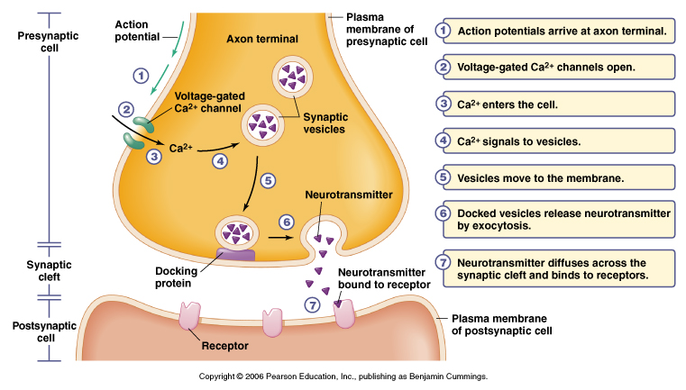
The sequence of events that lead to postsynaptic changes is as follows:
- The action potential signal arrives at the axon terminal (the bouton).
- The local depolarization causes Ca2+ channels to open. (Is this channel voltage, chemically, or mechanically gated? Answer.)
- Ca2+ enters the presynaptic cell because its concentration is greater outside the cell than inside.
- The Ca2+, by binding with calmodulin, causes vesicles filled with neurotransmitter to migrate towards the presynaptic membrane.
- The vesicle merges with the presynaptic membrane.
- The presynaptic membrane and vesicle now forms a continuous membrane, so that the neurotransmitter is released into the synaptic cleft. This process is called exocytosis.
- The neurotransmitter diffuses through the synaptic cleft and binds with receptor channel membranes that are located in both presynaptic and postsynaptic membranes. (Are these channels voltage, chemically, or mechanically gated? Answer.)
- The time period from neurotransmitter release to receptor channel binding is less than a millionth of a second.
There are two kinds of receptor channels: direct and indirect
- Direct: the receptor channel allows ions to pass through the membrane. The neurotransmitter acts like a key which opens the ion channel. This is the fastest kind of channel (about 0.5 ms). This is called an ionotropic receptor.
- Indirect: the binding of neurotransmitter to the receptor channel causes the release of a molecule, called a secondary messenger, that indirectly activates nearby ion channels. This is called a metabotropic receptor.
- This process is much slower than direct receptor ion channels: from 30 ms up to 1 second.
- However, this is the most common type of postsynaptic receptor channel
Once the postsynaptic ion channel is opened, whether directly or indirectly, the effect can be either excitatory (depolarizing) or inhibitory (hyperpolarizing).
- Excitatory Postsynaptic Potentials (EPSP)
- Excitatory ion channels are permeable to Na+ and K+
- Because of the electrical and concentration gradient, more Na+ moves into the cell than K+
- The inside of the cell becomes more positive, hence causing a local depolarization
- If enough depolarization occurs (for example, because the neurotransmitter released caused nearby ion channels to open), an action potential is generated
- Inhibitory Postsynaptic Potentials (IPSP)
- Inhibitory ion channels are permeable to Cl- and K+
- Because of the concentration gradient (not electrical), Cl- moves into the cell and K+ moves out of the cell
- The inside of the cell thus becomes more negative, hence causing a local hyperpolarization
- The hyperpolarization will make it more difficult for the cell membrane potential to reach threshold, thereby making it less likely that an action potential will be generated
- Depending on the kind of neurotransmitter released, the effect can be either excitatory or inhibitory
- The local excitatory depolarizations or inhibitory hyperpolarizations are graded (passive) potentials and therefore can summate or cause additive changes to the post-synaptic membrane potential. This process is known as summation
- Spatial summation occurs when multiple synapses in nearby locations are stimulated simultaneously
- Temporal summation occurs when the same channel is repeatedly opened (for example, because the presynaptic cell receives many impulses in a row), thereby altering the membrane potential further before it has the time to return to normal
- Although receptor ion channels are all chemically gated, enough depolarization past threshold can cause nearby voltage gated channels to open. An action potential would then be generated
If neurotransmitters were continually in the synaptic cleft, the postsynaptic channels would be continually stimulated and the membrane potential would not be able to become stable. There are three ways in which neurotransmitter is deactivated:
- Degradation: Enzymes located in the synaptic cleft break down the neurotransmitter into a substance which has no effect on the receptor channel
- Reuptake: The neurotransmitter can reenter the presynaptic cell through channels in the membrane.
- Autoreceptors: Receptors for a particular neurotransmitter are located on the presynaptic membrane that act like a thermostat. When there is too much neurotransmitter released in the synapse, it decreases the release of further neurotransmitter when the action potential arrives at the presynaptic membrane. It may accomplish this by decreasing the number of Ca2+ channels that open when the next action potential arrives at the presynaptic terminal
Neurotransmitters
A molecule is considered a neurotransmitter if it meets the following criteria:- Synthesis of the neurotransmitter occurs in the neuron itself
- It can be found in the presynaptic membrane (because it was carried there from the soma, or because it was synthesized there directly)
- Its release into the synaptic cleft causes a change in the postsynaptic membrane
- Its effect on a neuron is the same whether released exogenously (i.e., from outside the organism as a drug) or endogenously (from the presynaptic terminal)
- Once released, the molecule is specifically removed from the synaptic cleft either by reuse or degradation
- Small molecules, such as acetylcholine (ACh) or dopamine
- Are packaged in small vesicles
- Are released by exocytosis at active zones associated with Ca2+ channels
- Large molecules made up of chains of amino acids
- Are packaged in large vesicles (which can contain small molecules as well)
- Are released by exocytosis generally anywhere from the presynaptic membrane
Small Molecules
Acetylcholine (ACh)
- The only small molecule that is not an amino acid or derived from one
- Uses choline as a precursor
- Choline cannot be synthesized by the body and must be obtained from external food sources
- Used by motor neurons as an excitatory neurotransmitter in the spinal cord
- Used at neuromuscular junctions as an excitatory neurotransmitter to influence muscle activation
- Used by the Autonomic Nervous System, such as smooth muscles of the heart, as an inhibitory neurotransmitter in preganglionic neurons and postganglionic parasympathetic neurons
- Used everywhere in the brain. For example, memory systems of the CNS (may be related to Alzheimer's Disease).
- Most receptors for acetylcholine are ionotropic
Monoamines
a. Synthesized from tyrosine1. Dopamine

- Is synthesized in three steps from the amino acid tyrosine
- Is the direct precursor to norepinepherine.
- Enzyme converts tyrosine to L-DOPA
- Generally involved in regulatory motor activity
- In the basal ganglia, involved in mood, sensory perception, and attention
- Schizophrenics have too much dopamine, patients with Parkinson's Disease have too little

- Synthesized directly from dopamine, and forms the direct precursor to epinepherine. It is synthesized in four steps from tyrosine
- Synthesized within vesicles (the only neurotransmitter synthesized this way)
- Also known as noradrenaline
- Used in the CNS by neurons that project in the cortex, cerebellum, and spinal cord; as such has many uses including sleep/wakefulness regulation
- Activates sympathetic and parasympathetic neurons in the Autonomic Nervous System

- Synthesized in five steps from tyrosine, and directly from norepinepherine in the biosynthetic pathway
- Also known as adrenaline (from Latin: ad means "above" and renal means "kidney," while in Greek, epi means "above" and nephron means "kidney")
- Produced by the adrenal medulla, a gland above the kidney
- Few neurons in the brain use this neurotransmitter
- Activates sympathetic neurons in the Autonomic Nervous System
1. Serotonin (5-HT)

- Synthesized in two steps from the amino acid tryptophan
- Actual name: 5-hydroxytryptamine (5-HT)
- Regulates attention and other complex cognitive functions, such as sleep (dreaming), eating, mood, pain regulation
- Neurons which use serotonin are distributed throughout the brain and spinal cord
- Directly implicated in depression (also norepinepherine)
- Used by metabotropic receptors
Histamine

- Synthesized from the amino acid histidine
- Used in control of smooth muscle, exocrine glands, and vasculature
- High concentration in the hypothalamus, which regulates the secretion of horomones
- Used during inflammatory reactions
Amino Acids
Glutamate (Glu)
- Most prevalent neurotransmitter in the Central Nervous System. Used by more that 50% of neurons
- Derived from a -ketoglutarate
- Glutamate is the most important excitatory (EPSP) neurotransmitter, exciting about 90% of the postsynaptic terminals to which it contacts
- As an excitatory neutrotransmitter, it binds to with ionotropic receptors, causing depolarization by opening Na+ ion channels
- At metabotropic receptors, it is modulatory

- Synthesized directly from glutamate
- GABA is the most important inhibitory (IPSP) neurotransmitter
- Present in high concentrations in the CNS, preventing the brain from becoming overexcited
- As an inhibitory neutrotransmitter, it binds to both ionotropic and metabotropic receptors, causing hyperpolarization by opening Cl- ion channels
- Used by inhibitory interneurons in the spinal cord
Large Molecules
Neuropeptides- Derived from secretory proteins formed in the cell body
- They are first processed in the endoplasmic reticulum (ER) and are moved to the Golgi apparatus before being secreted as large vesicles and transported down the axon in preparation for exocytosis
- More than 50 peptides have been isolated in nerve cells. For example,
- Substance P and enkephalins: Active during inflammation and pain transmission in the PNS
- Endorphins: Endogenous opiates which cause euphoria, suppress pain, or regulate responses to stress
- Are either excitatory or inhibitory, and can also act as neuromodulators, affecting the amount of neurotransmitter released
- Some form part of the neuroendocrine system by functioning both as hormones and neurotransmitters
- Synthesis: Neurotransmitters are synthesized by the enzymatic transformation of precursors. The biosythetic pathway can be immediate (as in GABA from glutamate) or in multiple steps (as in epinepherine from norepinepherine from dopamine, etc.). The synthesis occurs either at the terminal boutons of the axon, or in the soma. In the latter case, it is transported to the axon terminals probably by way of microtubular tracks.
- Storage: They are packaged inside synaptic vesicles. These vesicles vary in size, depending on the size of the neurotransmitter.
- Release: The neurotransmitters are released from the presynaptic terminal by exocytosis and diffuse across the synaptic cleft to the postsynaptic membrane
- Binding: The neurotransmitters bind to receptor proteins imbedded in the postsynaptic cell's membrane. There are two kinds of receptors: ionotropic(direct) and metabotropic (indirect).
- Inactivation: The neurotransmitter is degraded either by being broken down enzymatically, or reused by active reuptake in which case the cycle begins again
Drugs
Drugs can affect any of the stages in the "life-cycle" of a neurotransmitter.Drugs that bind with receptors on the post-synaptic (and sometimes pre-synaptic) membrane fall into two groups:
- Agonists: Bind to receptors and simulate or enhance a neurotransmitter's actions (i.e., opening ion channels and causing EPSPs or IPSPs).
- Antagonists: Have the opposite effect of agonists by blocking the receptors and inactivating it (usually by taking up the space but without specifically causing the opening of the channel or the operation of the secondary messenger). The neurotransmitter's effect is nullified or diminished.
| Drug | Action (Brain) | Behavior |
| Nicotine | Acetylcholine receptor agonist | Smokers: relaxation, alertness, reduced desire for food. Non-smokers: Nausea, vomiting, cramps, and diarrhea. |
| Alcohol | 1. Reduces flow of Ca2+ into cells 2. GABA agonist 3. Increases number of binding sites for glutamate 4. Interferes with some secondary messenger systems | Low doses effect is excitatory. Moderate to high doses effect is inhibitory. |
| Cocaine and crack | Blocks reuptake of dopamine and norepinepherine | Feelings of well-being and confidence. Reduced desire for sleep and food. |
| Opiates (heroin, morphine, codeine) | Endorphin agonist | Pain suppression and euphoria. Suppresses cough and diarrhea |
| LSD | Serotonin receptor agonist | Visual hallucinations |
No comments:
Post a Comment