Thursday, 18 December 2014

Mutations


In genetics, a mutation is a permanent change of the nucleotide sequence of the genome of an organism, virus, or extrachromosomal genetic element. Mutations result from unrepaired damage to DNA or to RNA genomes (typically caused by radiation or chemical mutagens), errors in the process of replication, or from the insertion or deletion of segments of DNA by mobile genetic elements. Mutations may or may not produce discernible changes in the observable characteristics (phenotype) of an organism. Mutations play a part in both normal and abnormal biological processes including: evolution, cancer, and the development of the immune system.

Mutation can result in several different types of change in sequences. Mutations in genes can either have no effect, alter the product of a gene, or prevent the gene from functioning properly or completely. Mutations can also occur in nongenic regions. One study on genetic variations between different species of Drosophila suggests that, if a mutation changes a protein produced by a gene, the result is likely to be harmful, with an estimated 70 percent of amino acid polymorphisms that have damaging effects, and the remainder being either neutral or weakly beneficial. Due to the damaging effects that mutations can have on genes, organisms have mechanisms such as DNA repair to prevent or correct (revert the mutated sequence back to its original state) mutations

Myocardial Infarction


Myocardial infarction (MI; Latin: infarctus myocardii) or acute myocardial infarction (AMI), commonly known as a heart attack, occurs when blood stops flowing properly to a part of the heart, and the heart muscle is injured because it is not receiving enough oxygen. Usually, this is because one of the coronary arteries that supplies blood to the heart develops a blockage due to an unstable buildup of white blood cells, cholesterol and fat. The event is called "acute" if it is sudden and serious. Myocardial infarction differs from cardiac arrest, although cardiac arrest can be a consequence of MI.

A person having an acute MI usually has sudden chest pain that is felt behind the sternum and sometimes travels to the left arm or the left side of the neck. Additionally, the person may have shortness of breath, sweating, nausea, vomiting, abnormal heartbeats, and anxiety. Women experience fewer of these symptoms than men, but usually have shortness of breath, weakness, a feeling of indigestion, and fatigue. In many cases, in some estimates as high as 64%, the person does not have chest pain or has vague symptoms. These are called "silent" myocardial infarctions.

Important risks are previous cardiovascular disease, old age, tobacco smoking, abnormal blood levels of certain lipids, diabetes, high blood pressure, lack of physical activity, obesity, chronic kidney disease, excessive alcohol consumption, and the use of cocaine and amphetamines. The main ways to determine if a person has had a myocardial infarction are electrocardiograms (ECGs) that trace the electrical signals in the heart and testing the blood for substances associated with damage to the heart muscle. ECG testing is used to differentiate between two types of myocardial infarction based on the appearance of the tracing. An ST section of the tracing higher than the baseline is called an ST elevation MI (STEMI) which usually requires more aggressive treatment. If this is not the case, the diagnosis is confirmed with a blood test (usually troponin).

Immediate treatments for a suspected MI often include aspirin, which prevents further blood from clotting; nitroglycerin, sometimes given to treat chest pain; and oxygen. STEMI is treated by restoring circulation to the heart, called reperfusion therapy, and typical methods are angioplasty, where the arteries are pushed open, and thrombolysis, where the blockage is removed using medications. Non-ST elevation myocardial infarction (NSTEMI) may be managed with medication, although angioplasty may be required if the person is considered to be at high risk. who have multiple blockages of their coronary arteries, particularly if they also have diabetes, may also be treated with bypass surgery (CABG).Ischemic heart disease, which includes MI, angina, and heart failure when it happens after MI, was the leading cause of death for both men and women worldwide in 2011

Myofilament Contraction


Myofilaments are the filaments of myofibrils constructed from proteins. The principal types of muscle are striated muscle, obliquely striated muscle and smooth muscle. Various arrangements of myofilaments create different muscles. Striated muscle has transverse bands of filaments. In obliquely striated muscle, the filaments are staggered. Smooth muscle has irregular arrangements of filaments.

Muscle fiber contraction
The axon terminal of a motor neuron releases the neurotransmitter, acetylcholine.
Acetylcholine diffuses across the synaptic cleft and binds to the muscle fiber membrane.
This depolarizes the muscle fiber membrane, and the impulse travels to the muscle's sarcoplasmic reticulum via the transverse tubules.
Calcium ions are then released from the sarcoplasmic reticulum into the sarcoplasm and subsequently bind to troponin.
Troponin and the associated tropomyosin undergo a conformational change after calcium binding and expose the myosin binding sites on actin, the thin filament.
The filaments of actin and myosin then form linkages.
After binding, myosin pulls actin filaments toward each other, or inward.
Thus muscle contraction occurs, and the sarcomere shortens as this process takes place.
Myofilament.svg
Muscle fiber relaxation
The enzyme acetylcholinesterase breaks down acetylcholine and this ceases muscle fiber stimulation.
Active transport moves calcium ions back into the sarcoplasmic reticulum of the muscle fiber.
ATP causes the binding between actin and myosin filaments to break.
Troponin and tropomyosin revert to their original conformation and thereby block binding sites on the actin filament.
The muscle fiber relaxes and the entire sarcomere lengthens.
The muscle fiber is now prepared for the next contraction.

Olfaction Sense of Smell


Olfaction, also known as olfactics. is the sense of smell. This sense is mediated by specialized sensory cells of the nasal cavity of vertebrates, which can be considered analogous to sensory cells of the antennae of invertebrates. In humans, olfaction occurs when odorant molecules bind to specific sites on the olfactory receptors. These receptors are used to detect the presence of smell. They come together at the glomerulus, a structure which transmits signals to the olfactory bulb (a brain structure directly above the nasal cavity and below the frontal lobe). Many vertebrates, including most mammals and reptiles, have two distinct olfactory systems—the main olfactory system, and the accessory olfactory system (used mainly to detect pheromones). For air-breathing animals, the main olfactory system detects volatile chemicals, and the accessory olfactory system detects fluid-phase chemicals. Olfaction, along with taste, is a form of chemoreception. The chemicals themselves that activate the olfactory system, in general at very low concentrations, are called odorants. Although taste and smell are separate sensory systems in land animals, water-dwelling organisms often have one chemical sense.

Volatile small molecule odorants, non-volatile proteins, and non-volatile hydrocarbons may all produce olfactory sensations. Some animal species are able to smell carbon dioxide in minute concentrations


Oogenesis

Oogenesis, ovogenesis, or oögenesis /ˌoʊ.əˈdʒɛnɨsɪs/ is the creation of an ovum (egg cell). It is the female form of gametogenesis; the male equivalent is spermatogenesis. It involves the development of the various stages of the immature ovum

Oogenesis in mammals

Diagram showing the reduction in number of the chromosomes in the process of maturation of the ovum. (In mammals, the first polar body normally disintegrates before dividing, so only two polar bodies are produced.)
In mammals, the first part of oogenesis starts in the germinal epithelium, which gives rise to the development of ovarian follicles, the functional unit of the ovary.

Note that this process, important to all animal life cycles yet unlike all other instances of cell division, occurs completely without the aid of oo spindle-coordinating centrosomes.

Oogenesis consists of several sub-processes: oocytogenesis, ootidogenesis, and finally maturation to form an ovum (oogenesis proper). Folliculogenesis is a separate sub-process that accompanies and supports all three oogenetic sub-processes.

Cell type ploidy Process Process completion
Oogonium diploid Oocytogenesis (mitosis) third trimester (forming oocytes)
primary Oocyte diploid Ootidogenesis (meiosis 1) (Folliculogenesis) Dictyate in prophase I for up to 50 years
secondary Oocyte haploid Ootidogenesis (meiosis 2) Halted in metaphase II until fertilization
Ovum haploid
Oogonium —(Oocytogenesis)—> Primary Oocyte —(Meiosis I)—> First Polar Body (Discarded afterward) + Secondary oocyte —(Meiosis II)—> Second Polar Body (Discarded afterward) + Ovum

The creation of oogonia
The creation of oogonia traditionally doesn't belong to oogenesis proper, but, instead, to the common process of gametogenesis, which, in the female human, begins with the processes of folliculogenesis, oocytogenesis, and ootidogenesis.

Human oogenesis
At the start of the menstrual cycle, some 12-20 primary follicles begin to develop under the influence of elevated FSH to form secondary follicles. The primary follicles have formed from primordial follicles, which developed in the ovary at around 10–30 weeks after conception. By around day 9 of the cycle, only one healthy secondary follicle remains, with the rest having undergone ovarian follicle atresia. The remaining follicle is called the dominant follicle and is responsible for producing large amounts of estradiol during the late follicular phase. Estradiol production depends upon co-operation between the theca and granulosa cells. On day 14 of the cycle, an LH surge occurs, which itself is triggered by the positive feedback of estradiol. This causes the secondary follicle to develop into a tertiary follicle, which then ovulates some 24–36 hours later. An important event in the development of the tertiary follicle occurs when the primary oocyte completes the first meiotic division, resulting in the formation of a polar body and a secondary oocyte. The empty follicle then forms a corpus luteum, which later releases the hormone progesterone.

Oocytogenesis[edit]
Oogenesis starts with the process of developing oogonia, which occurs via the transformation of primordial follicles into primary oocytes, a process called oocytogenesis. Oocytogenesis is complete either before or shortly after birth.

Number of primary oocytes[edit]
It is commonly believed that, when oocytogenesis is complete, no additional primary oocytes are created, in contrast to the male process of spermatogenesis, where gametocytes are continuously created. In other words, primary oocytes reach their maximum development at ~20[5] weeks of gestational age, when approximately seven million primary oocytes have been created; however, at birth, this number has already been reduced to approximately 1-2 million.

Recently, however, two publications have challenged the belief that a finite number of oocytes are set around the time of birth.The renewal of ovarian follicles from germline stem cells (originating from bone marrow and peripheral blood) has been reported in the postnatal mouse ovary.

Due to the revolutionary nature of these claims, further experiments are required to determine the true dynamics of small follicle formation.

Ootidogenesis
The succeeding phase of ootidogenesis occurs when the primary oocyte develops into an ootid. This is achieved by the process of meiosis. In fact, a primary oocyte is, by its biological definition, a cell whose primary function is to divide by the process of meiosis.

However, although this process begins at prenatal age, it stops at prophase I. In late fetal life, all oocytes, still primary oocytes, have halted at this stage of development, called the dictyate. After menarche, these cells then continue to develop, although only a few do so every menstrual cycle.

Meiosis I
Meiosis I of ootidogenesis begins during embryonic development, but halts in the diplotene stage of prophase I until puberty. The mouse oocyte in the dictyate (prolonged diplotene) stage actively repairs DNA damage, whereas DNA repair is not detectable in the pre-dictyate (leptotene, zygotene and pachytene) stages of meiosis. For those primary oocytes that continue to develop in each menstrual cycle, however, synapsis occurs and tetrads form, enabling chromosomal crossover to occur. As a result of meiosis I, the primary oocyte has now developed into the secondary oocyte and the first polar body.

Meiosis II
Immediately after meiosis I, the haploid secondary oocyte initiates meiosis II. However, this process is also halted at the metaphase II stage until fertilization, if such should ever occur. When meiosis II has completed, an ootid and another polar body have now been created.

Folliculogenesis
Main article: Folliculogenesis
Synchronously with ootidogenesis, the ovarian follicle surrounding the ootid has developed from a primordial follicle to a preovulatory one.

Maturation into ovum
Both polar bodies disintegrate at the end of Meiosis II, leaving only the ootid, which then eventually undergoes maturation into a mature ovum.

The function of forming polar bodies is to discard the extra haploid sets of chromosomes that have resulted as a consequence of meiosis.

In vitro maturation
Main article: In vitro maturation
In vitro maturation (IVM) is the technique of letting ovarian follicles mature in vitro. It can potentially be performed before an IVF. In such cases, ovarian hyperstimulation isn't essential. Rather, oocytes can mature outside the body prior to IVF. Hence, no (or at least a lower dose of) gonadotropins have to be injected in the body. However, there still isn't enough evidence to prove the effectiveness and security of the technique.

Oogenesis in non-mammals
Main article: Evolution of sexual reproduction
Many protists produce egg cells in structures termed archegonia. Some algae and the oomycetes produce eggs in oogonia. In the brown alga Fucus, all four egg cells survive oogenesis, which is an exception to the rule that generally only one product of female meiosis survives to maturity.

In plants, oogenesis occurs inside the female gametophyte via mitosis. In many plants such as bryophytes, ferns, and gymnosperms, egg cells are formed in archegonia. In flowering plants, the female gametophyte has been reduced to an eight-celled embryo sac within the ovule inside the ovary of the flower. Oogenesis occurs within the embryo sac and leads to the formation of a single egg cell per ovule.

In ascaris, the oocyte does not even begin meiosis until the sperm touches it, in contrast to mammals, where meiosis is completed in the estrus cycle.

Organs of Digestion


In the human digestive system, the process of digestion has many stages, the first of which starts in the mouth (oral cavity). Digestion involves the breakdown of food into smaller and smaller components which can be absorbed and assimilated into the body. The secretion of saliva helps to produce a bolus which can be swallowed in the oesophagus to pass down into the stomach.

Saliva also contains a catalytic enzyme called amylase which starts to act on food in the mouth. Digestion is helped by the mastication of food by the teeth and also by the muscular contractions of peristalsis. Gastric juice in the stomach is essential for the continuation of digestion as is the production of mucus in the stomach.

Peristalsis is the rhythmic contraction of muscles that begins in the oesophagus and continues along the wall of the stomach and the rest of the gastrointestinal tract. This initially results in the production of chyme which when fully broken down in the small intestine is absorbed into the blood. Most of the digestion of food takes place in the small intestine. Water and some minerals are reabsorbed back into the blood, in the colon of the large intestine. The waste products of digestion are defecated from the anus via the rectum

Osteoporosis

Osteoporosis ("porous bones", from Greek: οστούν/ostoun meaning "bone" and πόρος/poros meaning "pore") is a progressive bone disease that is characterized by a decrease in bone mass and density which can lead to an increased risk of fracture.[1] In osteoporosis, the bone mineral density (BMD) is reduced, bone microarchitecture deteriorates, and the amount and variety of proteins in bone are altered. Osteoporosis is defined by the World Health Organization (WHO) as a bone mineral density of 2.5 standard deviations or more below the mean peak bone mass (average of young, healthy adults) as measured by dual-energy X-ray absorptiometry; the term "established osteoporosis" includes the presence of a fragility fracture.[2] The disease may be classified as primary type 1, primary type 2, or secondary. The form of osteoporosis most common in women after menopause is referred to as primary type 1 or postmenopausal osteoporosis, which is attributable to the decrease in estrogen production after menopause. Primary type 2 osteoporosis or senile osteoporosis occurs after age 75 and is seen in both females and males at a ratio of 2:1. Secondary osteoporosis may arise at any age and affect men and women equally; this form results from chronic predisposing medical problems or disease, or prolonged use of medications such as glucocorticoids, when the disease is called steroid- or glucocorticoid-induced osteoporosis.
The risk of osteoporosis fractures can be reduced with lifestyle changes and in those with previous osteoporosis related fractures, medications. Lifestyle change includes diet, exercise, and preventing falls. A review by the U.S. Preventive Services Task Force (USPSTF) found insufficient evidence to recommend calcium and vitamin D supplements to prevent fractures.[3] Bisphosphonates are useful in those with previous fractures from osteoporosis but are of minimal benefit in those who have osteoporosis but no previous fractures. Osteoporosis is a component of the frailty syndrome

Pain Pathway in Irritable Bowel Syndrome


Pancreas & Cystic Fibrosis


Cystic fibrosis (CF), also known as mucoviscidosis, is an autosomal recessive genetic disorder that affects mostly the lungs but also the pancreas, liver, and intestine. Difficulty breathing is the most serious symptom and results from frequent lung infections. Other symptoms—including sinus infections, poor growth, and infertility—affect other parts of the body.

CF is caused by one of many different mutations in the gene for the protein cystic fibrosis transmembrane conductance regulator (CFTR). This protein is required to regulate the components of sweat, digestive fluids, and mucus. Healthy people have two working copies of the CFTR gene. Carriers have one working copy. People with CF have no working copy. CF therefore has autosomal recessive inheritance. The underlying mechanism is abnormal transport of chloride and sodium across the epithelium, which is the cell layer that covers membranes over organs. This leads to thick, viscous secretions. Individuals with cystic fibrosis can be diagnosed before birth by genetic testing or by a sweat test in early childhood.

Lung infections are treated with antibiotics and other medications. Ultimately, lung transplantation is often necessary as CF worsens. The average life expectancy is 37 to 40 years in the United States. CF is most common among people of Central and Northern European ancestry, but occurs in many different groups around the world. It is rarest among Asians and the Middle Easterns.

The name cystic fibrosis refers to the characteristic scarring (fibrosis) and cyst formation within the pancreas, first recognized in the 1930s

Parathyroid Hormone

Parathyroid hormone (PTH), parathormone or parathyrin, is secreted by the chief cells of the parathyroid glands as a polypeptide containing 84 amino acids. It acts to increase the concentration of calcium (Ca2+) in the blood, whereas calcitonin (a hormone produced by the parafollicular cells (C cells) of the thyroid gland) acts to decrease calcium concentration. PTH acts to increase the concentration of calcium in the blood by acting upon the parathyroid hormone 1 receptor (high levels in bone and kidney) and the parathyroid hormone 2 receptor (high levels in the central nervous system, pancreas, testis, and placenta). PTH half-life is approximately 4 minutes. It has a molecular mass of 9.4 kDa

Penile Erection

An erection (clinically: penile erection or penile tumescence) is a physiological phenomenon in which the penis becomes firmer, engorged and enlarged. Penile erection is the result of a complex interaction of psychological, neural, vascular and endocrine factors, and is often associated with sexual arousal or sexual attraction, although erections can also be spontaneous. The shape, angle and direction of an erection varies considerably in humans.

Physiologically, erection is triggered by the parasympathetic division of the autonomic nervous system (ANS), causing nitric oxide (a vasodilator) levels to rise in the trabecular arteries and smooth muscle of the penis. The arteries dilate causing the corpora cavernosa of the penis (and to a lesser extent the corpora spongiosum) to fill with blood; simultaneously the ischiocavernosus and bulbospongiosus muscles compress the veins of the corpora cavernosa restricting the egress and circulation of this blood. Erection subsides when parasympathetic activity reduces to baseline.

As an autonomic nervous system response, an erection may result from a variety of stimuli, including sexual stimulation and sexual arousal, and is therefore not entirely under conscious control. Erections during sleep or upon waking up are known as nocturnal penile tumescence (NPT). Absence of nocturnal erection is commonly used to distinguish between physical and psychological causes of erectile dysfunction and impotence.Figure 28 01 06.jpg

A penis which is partly, but not fully, erect is sometimes known as a semi-erection (clinically: partial tumescence); a penis which is not erect is typically referred to as being flaccid, or soft.

Peptide Hormone Action


Peptide hormones are proteins that have an effect on the endocrine system of animals.

Like other proteins, peptide hormones are synthesized in cells from amino acids according to mRNA transcripts, which are synthesized from DNA templates inside the cell nucleus. Preprohormones, peptide hormone precursors, are then processed in several stages, typically in the endoplasmic reticulum, including removal of the N-terminal signal sequence and sometimes glycosylation, resulting in prohormones. The prohormones are then packaged into membrane-bound secretory vesicles, which can be secreted from the cell by exocytosis in response to specific stimuli (e.g. --an increase in Ca2+ and cAMP concentration in cytoplasm).

These prohormones often contain superfluous amino acid residues that were needed to direct folding of the hormone molecule into its active configuration but have no function once the hormone folds. Specific endopeptidases in the cell cleave the prohormone just before it is released into the bloodstream, generating the mature hormone form of the molecule. Mature peptide hormones then travel through the blood to all of the cells of the body, where they interact with specific receptors on the surfaces of their target cells. Some peptide/protein hormones (angiotensin II, basic fibroblast growth factor-2, parathyroid hormone-related protein) also interact with intracellular receptors located in the cytoplasm or nucleus by an intracrine mechanism.

Notable peptide hormones
Several important peptide hormones are secreted from the pituitary gland. The anterior pituitary secretes three: prolactin, which acts on the mammary gland; adrenocorticotropic hormone (ACTH), which acts on the adrenal cortex to regulate the secretion of glucocorticoids; and growth hormone, which acts on bone, muscle, and the liver. The posterior pituitary gland secretes antidiuretic hormone, also called vasopressin, and oxytocin. Peptide hormones are produced by many different organs and tissues, however, including the heart (atrial-natriuretic peptide (ANP) or atrial natriuretic factor (ANF)) and pancreas (glucagon, insulin and somatostatin), the gastrointestinal tract (cholecystokinin, gastrin), and adipose tissue stores (leptin).

Some neurotransmitters are secreted and released in a similar fashion to peptide hormones, and some 'neuropeptides' may be used as neurotransmitters in the nervous system in addition to acting as hormones when released into the blood. When a peptide hormone binds to receptors on the surface of the cell, a second messenger appears in the cytoplasm, which triggers intracellular responses

Photosynthesis Light Dependent


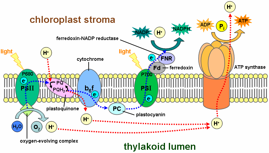
The light-dependent reactions take place on the thylakoid membranes. The inside of the thylakoid membrane is called the lumen, and outside the thylakoid membrane is the stroma, where the light-independent reactions take place. The thylakoid membrane contains some integral membrane protein complexes that catalyze the light reactions. There are four major protein complexes in the thylakoid membrane: Photosystem II (PSII), Cytochrome b6f complex, Photosystem I (PSI), and ATP synthase. These four complexes work together to ultimately create the products ATP and NADPH.
The two photosystems absorb light energy through pigments - primarily the chlorophylls, which are responsible for the green color of leaves. The light-dependent reactions begin in photosystem II. When a chlorophyll a molecule within the reaction center of PSII absorbs a photon, an electron in this molecule attains a higher energy level. Because this state of an electron is very unstable, the electron is transferred from one to another molecule creating a chain of redox reactions, called an electron transport chain (ETC). The electron flow goes from PSII to cytochrome b6f to PSI. In PSI, the electron gets the energy from another photon. The final electron acceptor is NADP. In oxygenic photosynthesis, the first electron donor is water, creating oxygen as a waste product. In anoxygenic photosynthesis various electron donors are used.

Cytochrome b6f and ATP synthase work together to create ATP. This process is called photophosphorylation, which occurs in two different ways. In non-cyclic photophosphorylation, cytochrome b6f uses the energy of electrons from PSII to pump protons from the stroma to the lumen. The proton gradient across the thylakoid membrane creates a proton-motive force, used by ATP synthase to form ATP. In cyclic photophosphorylation, cytochrome b6f uses the energy of electrons from not only PSII but also PSI to create more ATP and to stop the production of NADPH. Cyclic phosphorylation is important to create ATP and maintain NADPH in the right proportion for the light-independent reactions.

The net-reaction of all light-dependent reactions in oxygenic photosynthesis is:

2H
2O + 2NADP+
 + 3ADP + 3Pi → O
2 + 2NADPH + 3ATP

The two photosystems are protein complexes that absorb photons and are able to use this energy to create an electron transport chain. Photosystem I and II are very similar in structure and function. They use special proteins, called light-harvesting complexes, to absorb the photons with very high effectiveness. If a special pigment molecule in a photosynthetic reaction center absorbs a photon, an electron in this pigment attains the excited state and then is transferred to another molecule in the reaction center. This reaction, called photoinduced charge separation, is the start of the electron flow and is unique because it transforms light energy into chemical forms.

Phagocytic Cells

Phagocytic Cells

Phagocytes are cells that protect the body by ingesting (phagocytosing) harmful foreign particles, bacteria, and dead or dying cells. Their name comes from the Greek phagein, "to eat" or "devour", and "-cyte", the suffix in biology denoting "cell", from the Greek kutos, "hollow vessel". They are essential for fighting infections and for subsequent immunity. Phagocytes are important throughout the animal kingdom and are highly developed within vertebrates. One litre of human blood contains about six billion phagocytes. They were first discovered in 1882 by Ilya Ilyich Mechnikov while he was studying starfish larvae.Mechnikov was awarded the 1908 Nobel Prize in Physiology or Medicine for his discovery. Phagocytes occur in many species; some amoebae behave like macrophage phagocytes, which suggests that phagocytes appeared early in the evolution of life.

Phagocytes of humans and other animals are called "professional" or "non-professional" depending on how effective they are at phagocytosis.The professional phagocytes include many types of white blood cells (such as neutrophils, monocytes, macrophages, mast cells, and dendritic cells).The main difference between professional and non-professional phagocytes is that the professional phagocytes have molecules called receptors on their surfaces that can detect harmful objects, such as bacteria, that are not normally found in the body. Phagocytes are crucial in fighting infections, as well as in maintaining healthy tissues by removing dead and dying cells that have reached the end of their lifespan.

During an infection, chemical signals attract phagocytes to places where the pathogen has invaded the body. These chemicals may come from bacteria or from other phagocytes already present. The phagocytes move by a method called chemotaxis. When phagocytes come into contact with bacteria, the receptors on the phagocyte's surface will bind to them. This binding will lead to the engulfing of the bacteria by the phagocyte. Some phagocytes kill the ingested pathogen with oxidants and nitric oxide. After phagocytosis, macrophages and dendritic cells can also participate in antigen presentation, a process in which a phagocyte moves parts of the ingested material back to its surface. This material is then displayed to other cells of the immune system. Some phagocytes then travel to the body's lymph nodes and display the material to white blood cells called lymphocytes. This process is important in building immunity. However, many pathogens have evolved methods to evade attacks by phagocytes.

Photosynthesis Light Independent Reactions


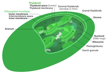
The light-independent reactions of photosynthesis are chemical reactions that convert carbon dioxide and other compounds into glucose. These reactions occur in the stroma, the fluid-filled area of a chloroplast outside of the thylakoid membranes. These reactions take the products(ATP and NADPH)of light-dependent reactions and perform further chemical processes on them. There are three phases to the light-independent reactions, collectively called the Calvin cycle: carbon fixation, reduction reactions, and ribulose 1,5-bisphosphate (RuBP) regeneration.
The internal structure of a chloroplast
Despite its name, this process occurs only when light is available. Plants do not carry out the Calvin cycle by night. They, instead, release sucrose into the phloem from their starch reserves. This process happens when light is available independent of the kind of photosynthesis (C3 carbon fixation, C4 carbon fixation, and Crassulacean Acid Metabolism); CAM plants store malic acid in their vacuoles every night and release it by day in order to make this process work.

Photosynthetic Electron Transport and ATP Synthesis


An electron transport chain (ETC) is a series of compounds that transfer electrons from electron donors to electron acceptors via redox reactions, and couples this electron transfer with the transfer of protons (H+ ions) across a membrane. This creates an electrochemical proton gradient that drives ATP synthesis, or the generation of chemical energy in the form of adenosine triphosphate (ATP). Final acceptor of electrons in electron transport chain is molecular oxygen.

Electron transport chains are used for extracting energy via redox reactions from sunlight in photosynthesis or, such as in the case of the oxidation of sugars, cellular respiration. In eukaryotes, an important electron transport chain is found in the inner mitochondrial membrane where it serves as the site of oxidative phosphorylation through the use of ATP synthase. It is also found in the thylakoid membrane of the chloroplast in photosynthetic eukaryotes. In bacteria, the electron transport chain is located in their cell membrane.

In chloroplasts, light drives the conversion of water to oxygen and NADP+ to NADPH with transfer of H+ ions across chloroplast membranes. In mitochondria, it is the conversion of oxygen to water, NADH to NAD+ and succinate to fumarate that are required to generate the proton gradient.

Electron transport chains are major sites of premature electron leakage to oxygen, generating superoxide and potentially resulting in increased oxidative stress

Wednesday, 17 December 2014

Phytochrome Signaling in Plants


Phytochrome signaling mechanisms and the control of plant development.
As they emerge from the ground, seedlings adopt a photosynthetic lifestyle, which is accompanied by dramatic changes in morphology and global alterations in gene expression that optimizes the plant body plan for light capture. Phytochromes are red and far-red photoreceptors that play a major role during photomorphogenesis, a complex developmental program that seedlings initiate when they first encounter light. The earliest phytochrome signaling events after excitation by red light include their rapid translocation from the cytoplasm to subnuclear bodies (photobodies) that contain other proteins involved in photomorphogenesis, including a number of transcription factors and E3 ligases. In the light, phytochromes and negatively acting transcriptional regulators that interact directly with phytochromes are destabilized, whereas positively acting transcriptional regulators are stabilized. Here, we discuss recent advances in our knowledge of the mechanisms linking phytochrome photoactivation in the cytoplasm and transcriptional regulation in the nucleus.

Polymerase Chain Reaction(detail)


The polymerase chain reaction (PCR) is a biomedical technology in molecular biology used to amplify a single copy or a few copies of a piece of DNA across several orders of magnitude, generating thousands to millions of copies of a particular DNA sequence.

Developed in 1983 by Kary Mullis, PCR is now a common and often indispensable technique used in medical and biological research labs for a variety of applications.These include DNA cloning for sequencing, DNA-based phylogeny, or functional analysis of genes; the diagnosis of hereditary diseases; the identification of genetic fingerprints (used in forensic sciences and paternity testing); and the detection and diagnosis of infectious diseases. In 1993, Mullis was awarded the Nobel Prize in Chemistry along with Michael Smith for his work on PCR.

The method relies on thermal cycling, consisting of cycles of repeated heating and cooling of the reaction for DNA melting and enzymatic replication of the DNA. Primers (short DNA fragments) containing sequences complementary to the target region along with a DNA polymerase, after which the method is named, are key components to enable selective and repeated amplification. As PCR progresses, the DNA generated is itself used as a template for replication, setting in motion a chain reaction in which the DNA template is exponentially amplified. PCR can be extensively modified to perform a wide array of genetic manipulations.

Almost all PCR applications employ a heat-stable DNA polymerase, such as Taq polymerase (an enzyme originally isolated from the bacterium Thermus aquaticus). This DNA polymerase enzymatically assembles a new DNA strand from DNA building-blocks, the nucleotides, by using single-stranded DNA as a template and DNA oligonucleotides (also called DNA primers), which are required for initiation of DNA synthesis. The vast majority of PCR methods use thermal cycling, i.e., alternately heating and cooling the PCR sample through a defined series of temperature steps.

In the first step, the two strands of the DNA double helix are physically separated at a high temperature in a process called DNA melting. In the second step, the temperature is lowered and the two DNA strands become templates for DNA polymerase to selectively amplify the target DNA. The selectivity of PCR results from the use of primers that are complementary to the DNA region targeted for amplification under specific thermal cycling conditions.
\


PCR principles and procedure


Figure 1a: A thermal cycler for PCR


Figure 1b: An older model three-temperature thermal cycler for PCR

PCR is used to amplify a specific region of a DNA strand (the DNA target). Most PCR methods typically amplify DNA fragments of between 0.1 and 10 kilo base pairs (kbp), although some techniques allow for amplification of fragments up to 40 kbp in size.[6]The amount of amplified product is determined by the available substrates in the reaction, which become limiting as the reaction progresses.[7]


A basic PCR set up requires several components and reagents.[8] These components include:


DNA template that contains the DNA region (target) to be amplified.

Two primers that are complementary to the 3' (three prime) ends of each of thesense and anti-sense strand of the DNA target.

Taq polymerase or another DNA polymerase with a temperature optimum at around 70 °C.

Deoxynucleoside triphosphates (dNTPs, sometimes called "deoxynucleotide triphosphates"; nucleotides containing triphosphate groups), the building-blocks from which the DNA polymerase synthesizes a new DNA strand.

Buffer solution, providing a suitable chemical environment for optimum activity and stability of the DNA polymerase.

Bivalent cations, magnesium or manganese ions; generally Mg2+ is used, but Mn2+ can be utilized for PCR-mediated DNA mutagenesis, as higher Mn2+concentration increases the error rate during DNA synthesis[9]

Monovalent cation potassium ions.

The PCR is commonly carried out in a reaction volume of 10–200 μl in small reaction tubes (0.2–0.5 ml volumes) in a thermal cycler. The thermal cycler heats and cools the reaction tubes to achieve the temperatures required at each step of the reaction (see below). Many modern thermal cyclers make use of the Peltier effect, which permits both heating and cooling of the block holding the PCR tubes simply by reversing the electric current. Thin-walled reaction tubes permit favorable thermal conductivity to allow for rapid thermal equilibration. Most thermal cyclers have heated lids to prevent condensation at the top of the reaction tube. Older thermocyclers lacking a heated lid require a layer of oil on top of the reaction mixture or a ball of wax inside the tube.


Procedure[edit]

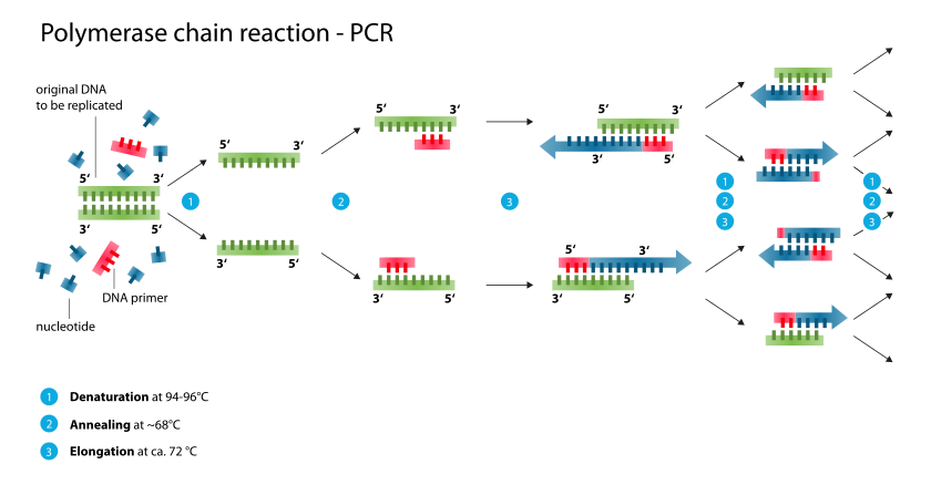
Typically, PCR consists of a series of 20-40 repeated temperature changes, called cycles, with each cycle commonly consisting of 2-3 discrete temperature steps, usually three (Figure below). The cycling is often preceded by a single temperature step at a high temperature (>90 °C), and followed by one hold at the end for final product extension or brief storage. The temperatures used and the length of time they are applied in each cycle depend on a variety of parameters. These include the enzyme used for DNA synthesis, the concentration of divalent ions and dNTPs in the reaction, and the melting temperature (Tm) of the primers.[10]


Initialization step(Only required for DNA polymerases that require heat activation by hot-start PCR.[11]): This step consists of heating the reaction to a temperature of 94–96 °C (or 98 °C if extremely thermostable polymerases are used), which is held for 1–9 minutes.

Denaturation step: This step is the first regular cycling event and consists of heating the reaction to 94–98 °C for 20–30 seconds. It causes DNA melting of the DNA template by disrupting the hydrogen bonds between complementary bases, yielding single-stranded DNA molecules.

Annealing step: The reaction temperature is lowered to 50–65 °C for 20–40 seconds allowing annealing of the primers to the single-stranded DNA template. This temperature needs to be low enough to allow for hybridization of the primer to the strand, but high enough in order for the hybridization to be specific, i.e. the primer should only bind to a perfectly complementary part of the template. If the temperature is too low, the primer could bind imperfectly. If it is too high, the primer might not bind. Typically the annealing temperature is about 3–5 °C below the Tm of the primers used. Stable DNA–DNA hydrogen bonds are only formed when the primer sequence very closely matches the template sequence. The polymerase binds to the primer-template hybrid and begins DNA formation.

Extension/elongation step: The temperature at this step depends on the DNA polymerase used; Taq polymerase has its optimum activity temperature at 75–80 °C,[12][13] and commonly a temperature of 72 °C is used with this enzyme. At this step the DNA polymerase synthesizes a new DNA strand complementary to the DNA template strand by adding dNTPs that are complementary to the template in 5' to 3' direction, condensing the 5'-phosphate group of the dNTPs with the 3'-hydroxyl group at the end of the nascent (extending) DNA strand. The extension time depends both on the DNA polymerase used and on the length of the DNA fragment to be amplified. As a rule-of-thumb, at its optimum temperature, the DNA polymerase will polymerize a thousand bases per minute. Under optimum conditions, i.e., if there are no limitations due to limiting substrates or reagents, at each extension step, the amount of DNA target is doubled, leading to exponential (geometric) amplification of the specific DNA fragment.

Final elongation: This single step is occasionally performed at a temperature of 70–74 °C (this is the temperature needed for optimal activity for most polymerases used in PCR) for 5–15 minutes after the last PCR cycle to ensure that any remaining single-stranded DNA is fully extended.

Final hold: This step at 4–15 °C for an indefinite time may be employed for short-term storage of the reaction.

Figure 2: Schematic drawing of the PCR cycle.

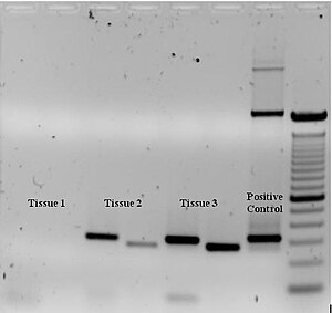
Figure 3: Ethidium bromide-stained PCR products after gel electrophoresis. Two sets of primers were used to amplify a target sequence from three different tissue samples. No amplification is present in sample #1; DNA bands in sample #2 and #3 indicate successful amplification of the target sequence. The gel also shows a positive control, and a DNA ladder containing DNA fragments of defined length for sizing the bands in the experimental PCRs.
To check whether the PCR generated the anticipated DNA fragment (also sometimes referred to as the amplimer or amplicon), agarose gel electrophoresis is employed for size separation of the PCR products. The size(s) of PCR products is determined by comparison with a DNA ladder (a molecular weight marker), which contains DNA fragments of known size, run on the gel alongside the PCR products (see Fig. 3).

PCR stages[edit]
The PCR process can be divided into three stages:

Exponential amplification: At every cycle, the amount of product is doubled (assuming 100% reaction efficiency). The reaction is very sensitive: only minute quantities of DNA need to be present.[14]

Leveling off stage: The reaction slows as the DNA polymerase loses activity and as consumption of reagents such as dNTPs and primers causes them to become limiting.

Plateau: No more product accumulates due to exhaustion of reagents and enzyme.

PCR optimization[edit]
Main article: PCR optimization
In practice, PCR can fail for various reasons, in part due to its sensitivity to contamination causing amplification of spurious DNA products. Because of this, a number of techniques and procedures have been developed for optimizing PCR conditions.[15][16] Contamination with extraneous DNA is addressed with lab protocols and procedures that separate pre-PCR mixtures from potential DNA contaminants.[8] This usually involves spatial separation of PCR-setup areas from areas for analysis or purification of PCR products, use of disposable plasticware, and thoroughly cleaning the work surface between reaction setups. Primer-design techniques are important in improving PCR product yield and in avoiding the formation of spurious products, and the usage of alternate buffer components or polymerase enzymes can help with amplification of long or otherwise problematic regions of DNA. Addition of reagents, such as formamide, in buffer systems may increase the specificity and yield of PCR.[17] Computer simulations of theoretical PCR results (Electronic PCR) may be performed to assist in primer design.[18]

Application of PCR[edit]
Main article: Applications of PCR
Selective DNA isolation[edit]
PCR allows isolation of DNA fragments from genomic DNA by selective amplification of a specific region of DNA. This use of PCR augments many methods, such as generating hybridization probes for Southern or northern hybridization and DNA cloning, which require larger amounts of DNA, representing a specific DNA region. PCR supplies these techniques with high amounts of pure DNA, enabling analysis of DNA samples even from very small amounts of starting material.

Other applications of PCR include DNA sequencing to determine unknown PCR-amplified sequences in which one of the amplification primers may be used in Sanger sequencing, isolation of a DNA sequence to expedite recombinant DNAtechnologies involving the insertion of a DNA sequence into a plasmid or the genetic material of another organism. Bacterial colonies (E. coli) can be rapidly screened by PCR for correct DNA vector constructs.[19] PCR may also be used for genetic fingerprinting; a forensic technique used to identify a person or organism by comparing experimental DNAs through different PCR-based methods.

Some PCR 'fingerprints' methods have high discriminative power and can be used to identify genetic relationships between individuals, such as parent-child or between siblings, and are used in paternity testing (Fig. 4). This technique may also be used to determine evolutionary relationships among organisms.[citation needed]

Figure 4: Electrophoresis of PCR-amplified DNA fragments. (1) Father. (2) Child. (3) Mother. The child has inherited some, but not all of the fingerprint of each of its parents, giving it a new, unique fingerprint.

Amplification and quantification of DNA[edit]

Because PCR amplifies the regions of DNA that it targets, PCR can be used to analyze extremely small amounts of sample. This is often critical for forensic analysis, when only a trace amount of DNA is available as evidence. PCR may also be used in the analysis of ADNA that is tens of thousands of years old. These PCR-based techniques have been successfully used on animals, such as a forty-thousand-year-old mammoth, and also on human DNA, in applications ranging from the analysis of Egyptianmummies to the identification of a Russian tsar and the body of English king Richard III.[20]


Quantitative PCR methods allow the estimation of the amount of a given sequence present in a sample—a technique often applied to quantitatively determine levels ofgene expression. Quantitative PCR is an established tool for DNA quantification that measures the accumulation of DNA product after each round of PCR amplification.


See also: Use of DNA in forensic entomology

PCR in diagnosis of diseases

PCR permits early diagnosis of malignant diseases such as leukemia and lymphomas, which is currently the highest-developed in cancer research and is already being used routinely. PCR assays can be performed directly on genomic DNA samples to detect translocation-specific malignant cells at a sensitivity that is at least 10,000 fold higher than that of other methods.
PCR allows for rapid and highly specific diagnosis of infectious diseases, including those caused by bacteria or virusesPCR also permits identification of non-cultivatable or slow-growing microorganisms such as mycobacteria, anaerobic bacteria, or viruses from tissue culture assays and animal models. The basis for PCR diagnostic applications in microbiology is the detection of infectious agents and the discrimination of non-pathogenic from pathogenic strains by virtue of specific genes.
Viral DNA can likewise be detected by PCR. The primers used need to be specific to the targeted sequences in the DNA of a virus, and the PCR can be used for diagnostic analyses or DNA sequencing of the viral genome. The high sensitivity of PCR permits virus detection soon after infection and even before the onset of disease. Such early detection may give physicians a significant lead time in treatment. The amount of virus ("viral load") in a patient can also be quantified by PCR-based DNA quantitation techniques (see below).

Variations on the basic PCR technique

  • Allele-specific PCR: a diagnostic or cloning technique based on single-nucleotide variations (SNVs not to be confused with SNPs) (single-base differences in a patient). It requires prior knowledge of a DNA sequence, including differences between alleles, and uses primers whose 3' ends encompass the SNV (base pair buffer around SNV usually incorporated). PCR amplification under stringent conditions is much less efficient in the presence of a mismatch between template and primer, so successful amplification with an SNP-specific primer signals presence of the specific SNP in a sequence.See SNP genotyping for more information.
  • Assembly PCR or Polymerase Cycling Assembly (PCA): artificial synthesis of long DNA sequences by performing PCR on a pool of long oligonucleotides with short overlapping segments. The oligonucleotides alternate between sense and antisense directions, and the overlapping segments determine the order of the PCR fragments, thereby selectively producing the final long DNA product.
  • Asymmetric PCR: preferentially amplifies one DNA strand in a double-stranded DNA template. It is used in sequencingand hybridization probing where amplification of only one of the two complementary strands is required. PCR is carried out as usual, but with a great excess of the primer for the strand targeted for amplification. Because of the slow (arithmetic) amplification later in the reaction after the limiting primer has been used up, extra cycles of PCR are required A recent modification on this process, known as Linear-After-The-Exponential-PCR (LATE-PCR), uses a limiting primer with a higher melting temperature (Tm) than the excess primer to maintain reaction efficiency as the limiting primer concentration decreases mid-reaction.
  • Dial-out PCR: a highly parallel method for retrieving accurate DNA molecules for gene synthesis. A complex library of DNA molecules is modified with unique flanking tags before massively parallel sequencing. Tag-directed primers then enable the retrieval of molecules with desired sequences by PCR.
  • Digital PCR (dPCR): used to measure the quantity of a target DNA sequence in a DNA sample. The DNA sample is highly diluted so that after running many PCRs in parallel, some of them will not receive a single molecule of the target DNA. The target DNA concentration is calculated using the proportion of negative outcomes. Hence the name 'digital PCR'.
  • Helicase-dependent amplification: similar to traditional PCR, but uses a constant temperature rather than cycling through denaturation and annealing/extension cycles. DNA helicase, an enzyme that unwinds DNA, is used in place of thermal denaturation.
  • Hot start PCR: a technique that reduces non-specific amplification during the initial set up stages of the PCR. It may be performed manually by heating the reaction components to the denaturation temperature (e.g., 95 °C) before adding the polymerase Specialized enzyme systems have been developed that inhibit the polymerase's activity at ambient temperature, either by the binding of an ] or by the presence of covalently bound inhibitors that dissociate only after a high-temperature activation step. Hot-start/cold-finish PCR is achieved with new hybrid polymerases that are inactive at ambient temperature and are instantly activated at elongation temperature.
  • In silico PCR (digital PCR, virtual PCR, electronic PCR, e-PCR) refers to computational tools used to calculate theoretical polymerase chain reaction results using a given set of primers (probes) to amplify DNA sequences from a sequenced genome or transcriptome. In silico PCR was proposed as an educational tool for molecular biology.
  • Intersequence-specific PCR (ISSR): a PCR method for DNA fingerprinting that amplifies regions between simple sequence repeats to produce a unique fingerprint of amplified fragment lengths.
  • Inverse PCR: is commonly used to identify the flanking sequences around genomic inserts. It involves a series of DNA digestions and self ligation, resulting in known sequences at either end of the unknown sequence.
  • Ligation-mediated PCR: uses small DNA linkers ligated to the DNA of interest and multiple primers annealing to the DNA linkers; it has been used for DNA sequencing, genome walking, and DNA footprinting.
  • Methylation-specific PCR (MSP): developed by Stephen Baylin and Jim Herman at the Johns Hopkins School of Medicine, and is used to detect methylation of CpG islands in genomic DNA. DNA is first treated with sodium bisulfite, which converts unmethylated cytosine bases to uracil, which is recognized by PCR primers as thymine. Two PCRs are then carried out on the modified DNA, using primer sets identical except at any CpG islands within the primer sequences. At these points, one primer set recognizes DNA with cytosines to amplify methylated DNA, and one set recognizes DNA with uracil or thymine to amplify unmethylated DNA. MSP using qPCR can also be performed to obtain quantitative rather than qualitative information about methylation.
  • Miniprimer PCR: uses a thermostable polymerase (S-Tbr) that can extend from short primers ("smalligos") as short as 9 or 10 nucleotides. This method permits PCR targeting to smaller primer binding regions, and is used to amplify conserved DNA sequences, such as the 16S (or eukaryotic 18S) rRNA gene.
  • Multiplex Ligation-dependent Probe Amplification (MLPA): permits multiple targets to be amplified with only a single primer pair, thus avoiding the resolution limitations of multiplex PCR (see below).
  • Multiplex-PCR: consists of multiple primer sets within a single PCR mixture to produce amplicons of varying sizes that are specific to different DNA sequences. By targeting multiple genes at once, additional information may be gained from a single test-run that otherwise would require several times the reagents and more time to perform. Annealing temperatures for each of the primer sets must be optimized to work correctly within a single reaction, and amplicon sizes. That is, their base pair length should be different enough to form distinct bands when visualized by gel electrophoresis.
  • Nanoparticle-Assisted PCR (nanoPCR): In recent years, it has been reported that some nanoparticles (NPs) can enhance the efficiency of PCR (thus being called nanoPCR), and some even perform better than the original PCR enhancers. It was also found that quantum dots (QDs) can improve PCR specificity and efficiency. Single-walled carbon nanotubes (SWCNTs) and multi-walled carbon nanotubes (MWCNTs) are efficient in enhancing the amplification of long PCR. Carbon nanopowder (CNP) was reported be able to improve the efficiency of repeated PCR and long PCR. ZnO, TiO2, and Ag NPs were also found to increase PCR yield. Importantly, already known data has indicated that non-metallic NPs retained acceptable amplification fidelity. Given that many NPs are capable of enhancing PCR efficiency, it is clear that there is likely to be great potential for nanoPCR technology improvements and product development.
  • Nested PCR: increases the specificity of DNA amplification, by reducing background due to non-specific amplification of DNA. Two sets of primers are used in two successive PCRs. In the first reaction, one pair of primers is used to generate DNA products, which besides the intended target, may still consist of non-specifically amplified DNA fragments. The product(s) are then used in a second PCR with a set of primers whose binding sites are completely or partially different from and located 3' of each of the primers used in the first reaction. Nested PCR is often more successful in specifically amplifying long DNA fragments than conventional PCR, but it requires more detailed knowledge of the target sequences.
  • Overlap-extension PCR or Splicing by overlap extension (SOEing) : a genetic engineering technique that is used to splice together two or more DNA fragments that contain complementary sequences. It is used to join DNA pieces containing genes, regulatory sequences, or mutations; the technique enables creation of specific and long DNA constructs. It can also introduce deletions, insertions or point mutations into a DNA sequence.
  • PAN-AC: uses isothermal conditions for amplification, and may be used in living cells.
  • quantitative PCR (qPCR): used to measure the quantity of a target sequence (commonly in real-time). It quantitatively measures starting amounts of DNA, cDNA, or RNA. quantitative PCR is commonly used to determine whether a DNA sequence is present in a sample and the number of its copies in the sample. Quantitative PCR has a very high degree of precision. Quantitative PCR methods use fluorescent dyes, such as Sybr Green, EvaGreen or fluorophore-containing DNA probes, such as TaqMan, to measure the amount of amplified product in real time. It is also sometimes abbreviated to RT-PCR (real-time PCR) but this abbreviation should be used only for reverse transcription PCR. qPCR is the appropriate contractions for quantitative PCR (real-time PCR).
  • Reverse Transcription PCR (RT-PCR): for amplifying DNA from RNA. Reverse transcriptase reverse transcribes RNAinto cDNA, which is then amplified by PCR. RT-PCR is widely used in expression profiling, to determine the expression of a gene or to identify the sequence of an RNA transcript, including transcription start and termination sites. If the genomic DNA sequence of a gene is known, RT-PCR can be used to map the location of exons and introns in the gene. The 5' end of a gene (corresponding to the transcription start site) is typically identified by RACE-PCR (Rapid Amplification of cDNA Ends).
  • Solid Phase PCR: encompasses multiple meanings, including Polony Amplification (where PCR colonies are derived in a gel matrix, for example), Bridge PCR (primers are covalently linked to a solid-support surface), conventional Solid Phase PCR (where Asymmetric PCR is applied in the presence of solid support bearing primer with sequence matching one of the aqueous primers) and Enhanced Solid Phase PCR(where conventional Solid Phase PCR can be improved by employing high Tm and nested solid support primer with optional application of a thermal 'step' to favour solid support priming).
  • Suicide PCR: typically used in paleogenetics or other studies where avoiding false positives and ensuring the specificity of the amplified fragment is the highest priority. It was originally described in a study to verify the presence of the microbe Yersinia pestis in dental samples obtained from 14th Century graves of people supposedly killed by plague during the medieval Black Death epidemic.[45] The method prescribes the use of any primer combination only once in a PCR (hence the term "suicide"), which should never have been used in any positive control PCR reaction, and the primers should always target a genomic region never amplified before in the lab using this or any other set of primers. This ensures that no contaminating DNA from previous PCR reactions is present in the lab, which could otherwise generate false positives.
  • Thermal asymmetric interlaced PCR (TAIL-PCR): for isolation of an unknown sequence flanking a known sequence. Within the known sequence, TAIL-PCR uses a nested pair of primers with differing annealing temperatures; a degenerate primer is used to amplify in the other direction from the unknown sequence.[46]
  • Touchdown PCR (Step-down PCR): a variant of PCR that aims to reduce nonspecific background by gradually lowering the annealing temperature as PCR cycling progresses. The annealing temperature at the initial cycles is usually a few degrees (3-5 °C) above the Tm of the primers used, while at the later cycles, it is a few degrees (3-5 °C) below the primer Tm. The higher temperatures give greater specificity for primer binding, and the lower temperatures permit more efficient amplification from the specific products formed during the initial cycles.
  • Universal Fast Walking: for genome walking and genetic fingerprinting using a more specific 'two-sided' PCR than conventional 'one-sided' approaches (using only one gene-specific primer and one general primer — which can lead to artefactual 'noise')by virtue of a mechanism involving lariat structure formation. Streamlined derivatives of UFW are LaNe RAGE (lariat-dependent nested PCR for rapid amplification of genomic DNA ends),5'RACE LaNe[ and 3'RACE LaNe.