Wednesday, 17 December 2014

Polymerase Chain Reaction

The polymerase chain reaction (PCR) is a biomedical technology in molecular biology used to amplify a single copy or a few copies of a piece of DNA across several orders of magnitude, generating thousands to millions of copies of a particular DNA sequence.

Developed in 1983 by Kary Mullis, PCR is now a common and often indispensable technique used in medical and biological research labs for a variety of applications. These include DNA cloning for sequencing, DNA-based phylogeny, or functional analysis of genes; the diagnosis of hereditary diseases; the identification of genetic fingerprints (used in forensic sciences and paternity testing); and the detection and diagnosis of infectious diseases. In 1993, Mullis was awarded the Nobel Prize in Chemistry along with Michael Smith for his work on PCR.

The method relies on thermal cycling, consisting of cycles of repeated heating and cooling of the reaction for DNA melting and enzymatic replication of the DNA. Primers (short DNA fragments) containing sequences complementary to the target region along with a DNA polymerase, after which the method is named, are key components to enable selective and repeated amplification. As PCR progresses, the DNA generated is itself used as a template for replication, setting in motion a chain reaction in which the DNA template is exponentially amplified. PCR can be extensively modified to perform a wide array of genetic manipulations.

Almost all PCR applications employ a heat-stable DNA polymerase, such as Taq polymerase (an enzyme originally isolated from the bacterium Thermus aquaticus). This DNA polymerase enzymatically assembles a new DNA strand from DNA building-blocks, the nucleotides, by using single-stranded DNA as a template and DNA oligonucleotides (also called DNA primers), which are required for initiation of DNA synthesis. The vast majority of PCR methods use thermal cycling, i.e., alternately heating and cooling the PCR sample through a defined series of temperature steps.

In the first step, the two strands of the DNA double helix are physically separated at a high temperature in a process called DNA melting. In the second step, the temperature is lowered and the two DNA strands become templates for DNA polymerase to selectively amplify the target DNA. The selectivity of PCR results from the use of primers that are complementary to the DNA region targeted for amplification under specific thermal cycling conditions.

Polyribosome


Polyribosomes (or polysomes) also known as ergosomes are a cluster of ribosomes, bound to a mRNA molecule, first discovered and characterized by Jonathan Warner, Paul Knopf, and Alex Rich in 1963. Many ribosomes read one mRNA simultaneously, progressing along the mRNA to synthesize the same protein. They may appear as clusters, linear polysomes, or circular rosettes on microscopy, but mainly circular in vivo. This circularization is aided by the fact that mRNA is able to be twisted into a circular formation, creating a cycle of rapid ribosome recycling, and utilization of ribosomes. The 5' 7-methylguanosine cap and 3' poly(A) tail present on eukaryotic mRNA aid in this process.

Polyribosomes can be found in three forms: free, cytoskeletal bound, and membrane bound.

Portal Venous System

In the circulatory system of animals, a portal venous system occurs when a capillary bed pools into another capillary bed through veins, without first going through the heart. Both capillary beds and the blood vessels that connect them are considered part of the portal venous system.

They are relatively uncommon as the majority of capillary beds drain into veins which then drain into the heart, not into another capillary bed. Portal venous systems are considered venous because the blood vessels that join the two capillary beds are either veins or venules.

Examples of such systems include the hepatic portal system, the hypophyseal portal system and (in non-mammals) the renal portal system. Unqualified, "portal venous system" often refers to the hepatic portal system. For this reason, "portal vein" most commonly refers to the hepatic portal vein.

The functional significance of such a system is that it transports products of one region directly to another region in relatively high concentrations. If the heart was involved in the blood circulation between those two regions, those products would be spread around the rest of the body.

In humans
The final common pathway for transport of venous blood from spleen, pancreas, gallbladder and the abdominal portion of the gastrointestinal tract (with the exception of the inferior part of the anal canal) is through the hepatic portal vein. The portal vein is formed by the union of the superior mesenteric vein and the splenic vein posterior to the neck of the pancreas at the level of vertebra body L1. Ascending towards the liver, the portal vein passes posterior to the superior part of the duodenum and enters the right margin of the lesser omentum, it is anterior to the omental foramen and posterior to both the bile duct, which is slightly to the right, and the hepatic artery proper, which is slightly to the left. On approaching the liver, the portal vein divides into right and left branches which enter the liver parenchyma. It gives tributaries; the right and left gastric veins, the cystic vein and the para-umbilical veins

Positive and Negative Feedback in the Body


Homeostasis

The biological definition of homeostasis is “the tendency of an organism or cell to regulate its internal environment and maintain equilibrium, usually by a system of feedback controls, so as to stabilize health and functioning”. Generally, the body is in homeostasis when it’s needs are met and it’s functioning properly.
Every organ in the body contributes to homeostasis. A complex set of chemical, thermal, and neural factors interact in complex ways, both helping and hindering the body while it works to maintain homeostasis.

Homeostatic control

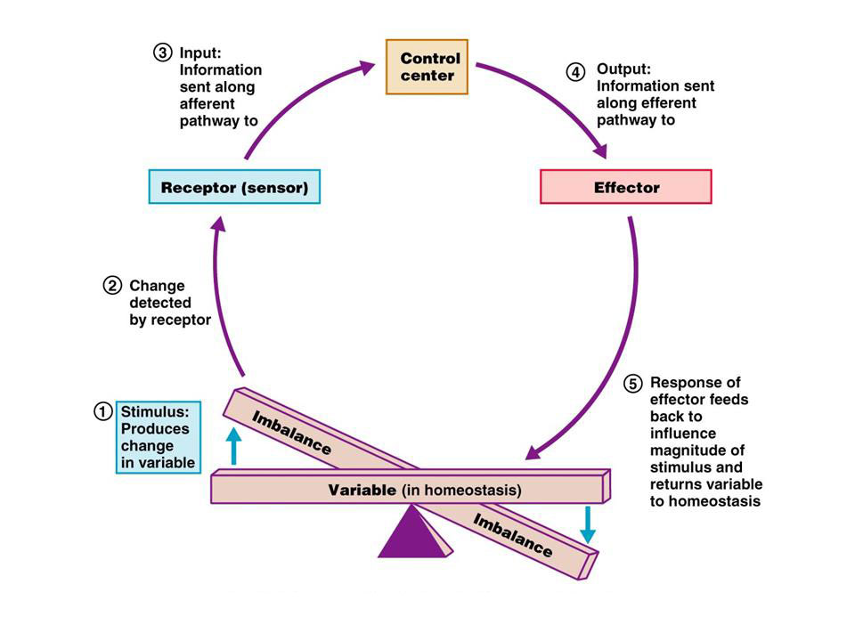
To maintain homeostasis, communication within the body is essential. The image below is an example of how a homeostatic control system works. Here is a brief explanation:
  1. Stimulus- produces a change to a variable (the factor being regulated).
  2. Receptor- detects the change. The receptor monitors the environment and responds to change (stimuli).
  3. Input- information travels along the (afferent) pathway to the control center. The control center determines the appropriate response and course of action.
  4. Output- information sent from the control center travels down the (efferent) pathway to the effector.
  5. Response- a response from the effector balances out the original stimulus to maintain homeostasis.
Image of a homeostatic control system using positive and negative feedback
Interactions among the elements of a homeostatic control system maintain stable internal conditions by using positive and negative feedback mechanisms.
Think of it as an extremely complex balancing act. Here’s a few more definitions you may want to know.
Afferent pathways- carry nerve impulses into the central nervous system. For instance, if you felt scorching heat on your hand, the message would travel through afferent pathways to your central nervous system.
Efferent pathways- carry nerve impulses away from the central nervous system to effectors (muscles, glands).
The feeling of heat would travel through an afferent pathway to the central nervous system. It would then interact with the effector and travel down the efferent pathway, eventually making the person remove their hand from the scorching heat.

Negative feedback mechanisms

Almost all homeostatic control mechanisms are negative feedback mechanisms. These mechanisms change the variable back to its original state or “ideal value”.
A good example of a negative feedback mechanism is a home thermostat (heating system). The thermostat contains the receptor (thermometer) and control center. If the heating system is set at 70 degrees Fahrenheit, the heat (effector) is turned on if the temperature drops below 70 degrees Fahrenheit. After the heater heats the house to 70 degrees Fahrenheit, it shuts off effectively maintaining the ideal temperature.
The control of blood sugar (glucose) by insulin is another good example of a negative feedback mechanism. When blood sugar rises, receptors in the body sense a change . In turn, the control center (pancreas) secretes insulin into the blood effectively lowering blood sugar levels. Once blood sugar levels reach homeostasis, the pancreas stops releasing insulin.
These are just two examples of negative feedback mechanisms within our body, there are 100’s, can you think of a few more?
Positive feedback mechanisms
A positive feedback mechanism is the exact opposite of a negative feedback mechanism. With negative feedback, the output reduces the original effect of the stimulus. In a positive feedback system, the output enhances the original stimulus. A good example of a positive feedback system is child birth. During labor, a hormone called oxytocin is released that intensifies and speeds up contractions. The increase in contractions causes more oxytocin to be released and the cycle goes on until the baby is born. The birth ends the release of oxytocin and ends the positive feedback mechanism.
Another good example of a positive feedback mechanism is blood clotting. Once a vessel is damaged, platelets start to cling to the injured site and release chemicals that attract more platelets. The platelets continue to pile up and release chemicals until a clot is formed.
Just remember that positive feedback mechanisms enhance the original stimulus and negative feedback mechanisms inhibit it.

Tuesday, 16 December 2014

Prion Diseases

A prion (Listeni/ˈpriːɒn/) is an infectious agent, specifically a protein in a misfolded form. The word prion, coined in 1982 by Stanley B. Prusiner, is derived from the words protein and infection. The protein itself, whether in its misfolded or its correctly folded form, can be referred to as the prion protein (PrP). A protein as an infectious agent stands in contrast to all other known infectious agents, like viruses, bacteria, fungi, or parasites—all of which must contain nucleic acids (either DNA, RNA, or both). Prions are responsible for mammalian transmissible spongiform encephalopathies, including bovine spongiform encephalopathy (BSE, also known as "mad cow disease") and scrapie in sheep. In humans, prions cause Creutzfeldt-Jakob Disease (CJD), variant Creutzfeldt-Jakob Disease (vCJD), Gerstmann–Sträussler–Scheinker syndrome, Fatal Familial Insomnia and kuru.[4] All known prion diseases in mammals affect the structure of the brain or other neural tissue and all are currently untreatable and universally fatal. In 2013, a study revealed that 1 in 2,000 people in the United Kingdom might harbour the infectious prion protein that causes vCJD.

Prions are not considered living organisms but are misfolded protein molecules which may propagate by transmitting a misfolded protein state. If a prion enters a healthy organism, it induces existing, properly folded proteins to convert into the disease-associated, misfolded prion form; the prion acts as a template to guide the misfolding of more proteins into prion form. These newly formed prions can then go on to convert more proteins themselves; this triggers a chain reaction that produces large amounts of the prion form. All known prions induce the formation of an amyloid fold, in which the protein polymerises into an aggregate consisting of tightly packed beta sheets. Amyloid aggregates are fibrils, growing at their ends, and replicating when breakage causes two growing ends to become four growing ends. The incubation period of prion diseases is determined by the exponential growth rate associated with prion replication, which is a balance between the linear growth and the breakage of aggregates. (Note that the propagation of the prion depends on the presence of normally folded protein in which the prion can induce misfolding; animals that do not express the normal form of the prion protein can neither develop nor transmit the disease.)

This altered structure is extremely stable and accumulates in infected tissue, causing tissue damage and cell death.[9] This structural stability means that prions are resistant to denaturation by chemical and physical agents, making disposal and containment of these particles difficult. Prions come in different strains, each with a slightly different structure, and, most of the time, strains breed true. Prion replication is nevertheless subject to occasional epimutation and then natural selection just like other forms of replication.

All known mammalian prion diseases are caused by the so-called prion protein, PrP. The endogenous, properly folded form is denoted PrPC (for Common or Cellular), whereas the disease-linked, misfolded form is denoted PrPSc (for Scrapie, after one of the diseases first linked to prions and neurodegeneration.)[11][12] The precise structure of the prion is not known, though they can be formed by combining PrPC, polyadenylic acid, and lipids in a Protein Misfolding Cyclic Amplification (PMCA) reaction. Proteins showing prion-type behavior are also found in some fungi, which has been useful in helping to understand mammalian prions. Fungal prions do not appear to cause disease in their hosts

Processing of Gene Information Prokaryotes versus Eukaryotes

Prokaryotic versus Eukaryotic Gene Expression

To understand how gene expression is regulated, we must first understand how a gene codes for a functional protein in a cell. The process occurs in both prokaryotic and eukaryotic cells, just in slightly different manners.
Prokaryotic organisms are single-celled organisms that lack a defined nucleus; therefore, their DNA floats freely within the cell cytoplasm. To synthesize a protein, the processes of transcription (DNA to RNA) and translation (RNA to protein) occur almost simultaneously. When the resulting protein is no longer needed, transcription stops. Thus, the regulation of transcription is the primary method to control what type of protein and how much of each protein is expressed in a prokaryotic cell. All of the subsequent steps occur automatically. When more protein is required, more transcription occurs. Therefore, in prokaryotic cells, the control of gene expression is mostly at the transcriptional level.
Eukaryotic cells, in contrast, have intracellular organelles that add to their complexity. In eukaryotic cells, the DNA is contained inside the cell's nucleus where it is transcribed into RNA. The newly-synthesized RNA is then transported out of the nucleus into the cytoplasm where ribosomes translate the RNA into protein. The processes of transcription and translation are physically separated by the nuclear membrane; transcription occurs only within the nucleus, and translation occurs only outside the nucleus within the cytoplasm. The regulation of gene expression can occur at all stages of the process . Regulation may occur when the DNA is uncoiled and loosened from nucleosomes to bind transcription factors (epigenetics), when the RNA is transcribed (transcriptional level), when the RNA is processed and exported to the cytoplasm after it is transcribed (post-transcriptional level), when the RNA is translated into protein (translational level), or after the protein has been made (post-translational level).
Prokaryotic vs Eukaryotic Gene Expression
Prokaryotic transcription and translation occur simultaneously in the cytoplasm, and regulation occurs at the transcriptional level. Eukaryotic gene expression is regulated during transcription and RNA processing, which take place in the nucleus, and during protein translation, which takes place in the cytoplasm. Further regulation may occur through post-translational modifications of proteins.


Protein Structure and Protein Denaturation

Denaturation is a process in which proteins or nucleic acids lose the quaternary structure, tertiary structure and secondary structure which is present in their native state, by application of some external stress or compound such as a strong acid or base, a concentrated inorganic salt, an organic solvent (e.g., alcohol or chloroform), radiation or heat. If proteins in a living cell are denatured, this results in disruption of cell activity and possibly cell death. Denatured proteins can exhibit a wide range of characteristics, from conformational change and loss of solubility to communal aggregation.

This concept is unrelated to denatured alcohol, which is alcohol that has been mixed with additives to make it unsuitable for human consumption.