Showing posts with label 3D-MEDICAL VEDIO. Show all posts

Thursday, 18 December 2014

Myocardial Infarction


Myocardial infarction (MI; Latin: infarctus myocardii) or acute myocardial infarction (AMI), commonly known as a heart attack, occurs when blood stops flowing properly to a part of the heart, and the heart muscle is injured because it is not receiving enough oxygen. Usually, this is because one of the coronary arteries that supplies blood to the heart develops a blockage due to an unstable buildup of white blood cells, cholesterol and fat. The event is called "acute" if it is sudden and serious. Myocardial infarction differs from cardiac arrest, although cardiac arrest can be a consequence of MI.

A person having an acute MI usually has sudden chest pain that is felt behind the sternum and sometimes travels to the left arm or the left side of the neck. Additionally, the person may have shortness of breath, sweating, nausea, vomiting, abnormal heartbeats, and anxiety. Women experience fewer of these symptoms than men, but usually have shortness of breath, weakness, a feeling of indigestion, and fatigue. In many cases, in some estimates as high as 64%, the person does not have chest pain or has vague symptoms. These are called "silent" myocardial infarctions.

Important risks are previous cardiovascular disease, old age, tobacco smoking, abnormal blood levels of certain lipids, diabetes, high blood pressure, lack of physical activity, obesity, chronic kidney disease, excessive alcohol consumption, and the use of cocaine and amphetamines. The main ways to determine if a person has had a myocardial infarction are electrocardiograms (ECGs) that trace the electrical signals in the heart and testing the blood for substances associated with damage to the heart muscle. ECG testing is used to differentiate between two types of myocardial infarction based on the appearance of the tracing. An ST section of the tracing higher than the baseline is called an ST elevation MI (STEMI) which usually requires more aggressive treatment. If this is not the case, the diagnosis is confirmed with a blood test (usually troponin).

Immediate treatments for a suspected MI often include aspirin, which prevents further blood from clotting; nitroglycerin, sometimes given to treat chest pain; and oxygen. STEMI is treated by restoring circulation to the heart, called reperfusion therapy, and typical methods are angioplasty, where the arteries are pushed open, and thrombolysis, where the blockage is removed using medications. Non-ST elevation myocardial infarction (NSTEMI) may be managed with medication, although angioplasty may be required if the person is considered to be at high risk. who have multiple blockages of their coronary arteries, particularly if they also have diabetes, may also be treated with bypass surgery (CABG).Ischemic heart disease, which includes MI, angina, and heart failure when it happens after MI, was the leading cause of death for both men and women worldwide in 2011

Myofilament Contraction


Myofilaments are the filaments of myofibrils constructed from proteins. The principal types of muscle are striated muscle, obliquely striated muscle and smooth muscle. Various arrangements of myofilaments create different muscles. Striated muscle has transverse bands of filaments. In obliquely striated muscle, the filaments are staggered. Smooth muscle has irregular arrangements of filaments.

Muscle fiber contraction
The axon terminal of a motor neuron releases the neurotransmitter, acetylcholine.
Acetylcholine diffuses across the synaptic cleft and binds to the muscle fiber membrane.
This depolarizes the muscle fiber membrane, and the impulse travels to the muscle's sarcoplasmic reticulum via the transverse tubules.
Calcium ions are then released from the sarcoplasmic reticulum into the sarcoplasm and subsequently bind to troponin.
Troponin and the associated tropomyosin undergo a conformational change after calcium binding and expose the myosin binding sites on actin, the thin filament.
The filaments of actin and myosin then form linkages.
After binding, myosin pulls actin filaments toward each other, or inward.
Thus muscle contraction occurs, and the sarcomere shortens as this process takes place.
Myofilament.svg
Muscle fiber relaxation
The enzyme acetylcholinesterase breaks down acetylcholine and this ceases muscle fiber stimulation.
Active transport moves calcium ions back into the sarcoplasmic reticulum of the muscle fiber.
ATP causes the binding between actin and myosin filaments to break.
Troponin and tropomyosin revert to their original conformation and thereby block binding sites on the actin filament.
The muscle fiber relaxes and the entire sarcomere lengthens.
The muscle fiber is now prepared for the next contraction.

Olfaction Sense of Smell


Olfaction, also known as olfactics. is the sense of smell. This sense is mediated by specialized sensory cells of the nasal cavity of vertebrates, which can be considered analogous to sensory cells of the antennae of invertebrates. In humans, olfaction occurs when odorant molecules bind to specific sites on the olfactory receptors. These receptors are used to detect the presence of smell. They come together at the glomerulus, a structure which transmits signals to the olfactory bulb (a brain structure directly above the nasal cavity and below the frontal lobe). Many vertebrates, including most mammals and reptiles, have two distinct olfactory systems—the main olfactory system, and the accessory olfactory system (used mainly to detect pheromones). For air-breathing animals, the main olfactory system detects volatile chemicals, and the accessory olfactory system detects fluid-phase chemicals. Olfaction, along with taste, is a form of chemoreception. The chemicals themselves that activate the olfactory system, in general at very low concentrations, are called odorants. Although taste and smell are separate sensory systems in land animals, water-dwelling organisms often have one chemical sense.

Volatile small molecule odorants, non-volatile proteins, and non-volatile hydrocarbons may all produce olfactory sensations. Some animal species are able to smell carbon dioxide in minute concentrations


Oogenesis

Oogenesis, ovogenesis, or oögenesis /ˌoʊ.əˈdʒɛnɨsɪs/ is the creation of an ovum (egg cell). It is the female form of gametogenesis; the male equivalent is spermatogenesis. It involves the development of the various stages of the immature ovum

Oogenesis in mammals

Diagram showing the reduction in number of the chromosomes in the process of maturation of the ovum. (In mammals, the first polar body normally disintegrates before dividing, so only two polar bodies are produced.)
In mammals, the first part of oogenesis starts in the germinal epithelium, which gives rise to the development of ovarian follicles, the functional unit of the ovary.

Note that this process, important to all animal life cycles yet unlike all other instances of cell division, occurs completely without the aid of oo spindle-coordinating centrosomes.

Oogenesis consists of several sub-processes: oocytogenesis, ootidogenesis, and finally maturation to form an ovum (oogenesis proper). Folliculogenesis is a separate sub-process that accompanies and supports all three oogenetic sub-processes.

Cell type ploidy Process Process completion
Oogonium diploid Oocytogenesis (mitosis) third trimester (forming oocytes)
primary Oocyte diploid Ootidogenesis (meiosis 1) (Folliculogenesis) Dictyate in prophase I for up to 50 years
secondary Oocyte haploid Ootidogenesis (meiosis 2) Halted in metaphase II until fertilization
Ovum haploid
Oogonium —(Oocytogenesis)—> Primary Oocyte —(Meiosis I)—> First Polar Body (Discarded afterward) + Secondary oocyte —(Meiosis II)—> Second Polar Body (Discarded afterward) + Ovum

The creation of oogonia
The creation of oogonia traditionally doesn't belong to oogenesis proper, but, instead, to the common process of gametogenesis, which, in the female human, begins with the processes of folliculogenesis, oocytogenesis, and ootidogenesis.

Human oogenesis
At the start of the menstrual cycle, some 12-20 primary follicles begin to develop under the influence of elevated FSH to form secondary follicles. The primary follicles have formed from primordial follicles, which developed in the ovary at around 10–30 weeks after conception. By around day 9 of the cycle, only one healthy secondary follicle remains, with the rest having undergone ovarian follicle atresia. The remaining follicle is called the dominant follicle and is responsible for producing large amounts of estradiol during the late follicular phase. Estradiol production depends upon co-operation between the theca and granulosa cells. On day 14 of the cycle, an LH surge occurs, which itself is triggered by the positive feedback of estradiol. This causes the secondary follicle to develop into a tertiary follicle, which then ovulates some 24–36 hours later. An important event in the development of the tertiary follicle occurs when the primary oocyte completes the first meiotic division, resulting in the formation of a polar body and a secondary oocyte. The empty follicle then forms a corpus luteum, which later releases the hormone progesterone.

Oocytogenesis[edit]
Oogenesis starts with the process of developing oogonia, which occurs via the transformation of primordial follicles into primary oocytes, a process called oocytogenesis. Oocytogenesis is complete either before or shortly after birth.

Number of primary oocytes[edit]
It is commonly believed that, when oocytogenesis is complete, no additional primary oocytes are created, in contrast to the male process of spermatogenesis, where gametocytes are continuously created. In other words, primary oocytes reach their maximum development at ~20[5] weeks of gestational age, when approximately seven million primary oocytes have been created; however, at birth, this number has already been reduced to approximately 1-2 million.

Recently, however, two publications have challenged the belief that a finite number of oocytes are set around the time of birth.The renewal of ovarian follicles from germline stem cells (originating from bone marrow and peripheral blood) has been reported in the postnatal mouse ovary.

Due to the revolutionary nature of these claims, further experiments are required to determine the true dynamics of small follicle formation.

Ootidogenesis
The succeeding phase of ootidogenesis occurs when the primary oocyte develops into an ootid. This is achieved by the process of meiosis. In fact, a primary oocyte is, by its biological definition, a cell whose primary function is to divide by the process of meiosis.

However, although this process begins at prenatal age, it stops at prophase I. In late fetal life, all oocytes, still primary oocytes, have halted at this stage of development, called the dictyate. After menarche, these cells then continue to develop, although only a few do so every menstrual cycle.

Meiosis I
Meiosis I of ootidogenesis begins during embryonic development, but halts in the diplotene stage of prophase I until puberty. The mouse oocyte in the dictyate (prolonged diplotene) stage actively repairs DNA damage, whereas DNA repair is not detectable in the pre-dictyate (leptotene, zygotene and pachytene) stages of meiosis. For those primary oocytes that continue to develop in each menstrual cycle, however, synapsis occurs and tetrads form, enabling chromosomal crossover to occur. As a result of meiosis I, the primary oocyte has now developed into the secondary oocyte and the first polar body.

Meiosis II
Immediately after meiosis I, the haploid secondary oocyte initiates meiosis II. However, this process is also halted at the metaphase II stage until fertilization, if such should ever occur. When meiosis II has completed, an ootid and another polar body have now been created.

Folliculogenesis
Main article: Folliculogenesis
Synchronously with ootidogenesis, the ovarian follicle surrounding the ootid has developed from a primordial follicle to a preovulatory one.

Maturation into ovum
Both polar bodies disintegrate at the end of Meiosis II, leaving only the ootid, which then eventually undergoes maturation into a mature ovum.

The function of forming polar bodies is to discard the extra haploid sets of chromosomes that have resulted as a consequence of meiosis.

In vitro maturation
Main article: In vitro maturation
In vitro maturation (IVM) is the technique of letting ovarian follicles mature in vitro. It can potentially be performed before an IVF. In such cases, ovarian hyperstimulation isn't essential. Rather, oocytes can mature outside the body prior to IVF. Hence, no (or at least a lower dose of) gonadotropins have to be injected in the body. However, there still isn't enough evidence to prove the effectiveness and security of the technique.

Oogenesis in non-mammals
Main article: Evolution of sexual reproduction
Many protists produce egg cells in structures termed archegonia. Some algae and the oomycetes produce eggs in oogonia. In the brown alga Fucus, all four egg cells survive oogenesis, which is an exception to the rule that generally only one product of female meiosis survives to maturity.

In plants, oogenesis occurs inside the female gametophyte via mitosis. In many plants such as bryophytes, ferns, and gymnosperms, egg cells are formed in archegonia. In flowering plants, the female gametophyte has been reduced to an eight-celled embryo sac within the ovule inside the ovary of the flower. Oogenesis occurs within the embryo sac and leads to the formation of a single egg cell per ovule.

In ascaris, the oocyte does not even begin meiosis until the sperm touches it, in contrast to mammals, where meiosis is completed in the estrus cycle.

Organs of Digestion


In the human digestive system, the process of digestion has many stages, the first of which starts in the mouth (oral cavity). Digestion involves the breakdown of food into smaller and smaller components which can be absorbed and assimilated into the body. The secretion of saliva helps to produce a bolus which can be swallowed in the oesophagus to pass down into the stomach.

Saliva also contains a catalytic enzyme called amylase which starts to act on food in the mouth. Digestion is helped by the mastication of food by the teeth and also by the muscular contractions of peristalsis. Gastric juice in the stomach is essential for the continuation of digestion as is the production of mucus in the stomach.

Peristalsis is the rhythmic contraction of muscles that begins in the oesophagus and continues along the wall of the stomach and the rest of the gastrointestinal tract. This initially results in the production of chyme which when fully broken down in the small intestine is absorbed into the blood. Most of the digestion of food takes place in the small intestine. Water and some minerals are reabsorbed back into the blood, in the colon of the large intestine. The waste products of digestion are defecated from the anus via the rectum

Osteoporosis

Osteoporosis ("porous bones", from Greek: οστούν/ostoun meaning "bone" and πόρος/poros meaning "pore") is a progressive bone disease that is characterized by a decrease in bone mass and density which can lead to an increased risk of fracture.[1] In osteoporosis, the bone mineral density (BMD) is reduced, bone microarchitecture deteriorates, and the amount and variety of proteins in bone are altered. Osteoporosis is defined by the World Health Organization (WHO) as a bone mineral density of 2.5 standard deviations or more below the mean peak bone mass (average of young, healthy adults) as measured by dual-energy X-ray absorptiometry; the term "established osteoporosis" includes the presence of a fragility fracture.[2] The disease may be classified as primary type 1, primary type 2, or secondary. The form of osteoporosis most common in women after menopause is referred to as primary type 1 or postmenopausal osteoporosis, which is attributable to the decrease in estrogen production after menopause. Primary type 2 osteoporosis or senile osteoporosis occurs after age 75 and is seen in both females and males at a ratio of 2:1. Secondary osteoporosis may arise at any age and affect men and women equally; this form results from chronic predisposing medical problems or disease, or prolonged use of medications such as glucocorticoids, when the disease is called steroid- or glucocorticoid-induced osteoporosis.
The risk of osteoporosis fractures can be reduced with lifestyle changes and in those with previous osteoporosis related fractures, medications. Lifestyle change includes diet, exercise, and preventing falls. A review by the U.S. Preventive Services Task Force (USPSTF) found insufficient evidence to recommend calcium and vitamin D supplements to prevent fractures.[3] Bisphosphonates are useful in those with previous fractures from osteoporosis but are of minimal benefit in those who have osteoporosis but no previous fractures. Osteoporosis is a component of the frailty syndrome

Pain Pathway in Irritable Bowel Syndrome


Pancreas & Cystic Fibrosis


Cystic fibrosis (CF), also known as mucoviscidosis, is an autosomal recessive genetic disorder that affects mostly the lungs but also the pancreas, liver, and intestine. Difficulty breathing is the most serious symptom and results from frequent lung infections. Other symptoms—including sinus infections, poor growth, and infertility—affect other parts of the body.

CF is caused by one of many different mutations in the gene for the protein cystic fibrosis transmembrane conductance regulator (CFTR). This protein is required to regulate the components of sweat, digestive fluids, and mucus. Healthy people have two working copies of the CFTR gene. Carriers have one working copy. People with CF have no working copy. CF therefore has autosomal recessive inheritance. The underlying mechanism is abnormal transport of chloride and sodium across the epithelium, which is the cell layer that covers membranes over organs. This leads to thick, viscous secretions. Individuals with cystic fibrosis can be diagnosed before birth by genetic testing or by a sweat test in early childhood.

Lung infections are treated with antibiotics and other medications. Ultimately, lung transplantation is often necessary as CF worsens. The average life expectancy is 37 to 40 years in the United States. CF is most common among people of Central and Northern European ancestry, but occurs in many different groups around the world. It is rarest among Asians and the Middle Easterns.

The name cystic fibrosis refers to the characteristic scarring (fibrosis) and cyst formation within the pancreas, first recognized in the 1930s

Parathyroid Hormone

Parathyroid hormone (PTH), parathormone or parathyrin, is secreted by the chief cells of the parathyroid glands as a polypeptide containing 84 amino acids. It acts to increase the concentration of calcium (Ca2+) in the blood, whereas calcitonin (a hormone produced by the parafollicular cells (C cells) of the thyroid gland) acts to decrease calcium concentration. PTH acts to increase the concentration of calcium in the blood by acting upon the parathyroid hormone 1 receptor (high levels in bone and kidney) and the parathyroid hormone 2 receptor (high levels in the central nervous system, pancreas, testis, and placenta). PTH half-life is approximately 4 minutes. It has a molecular mass of 9.4 kDa

Penile Erection

An erection (clinically: penile erection or penile tumescence) is a physiological phenomenon in which the penis becomes firmer, engorged and enlarged. Penile erection is the result of a complex interaction of psychological, neural, vascular and endocrine factors, and is often associated with sexual arousal or sexual attraction, although erections can also be spontaneous. The shape, angle and direction of an erection varies considerably in humans.

Physiologically, erection is triggered by the parasympathetic division of the autonomic nervous system (ANS), causing nitric oxide (a vasodilator) levels to rise in the trabecular arteries and smooth muscle of the penis. The arteries dilate causing the corpora cavernosa of the penis (and to a lesser extent the corpora spongiosum) to fill with blood; simultaneously the ischiocavernosus and bulbospongiosus muscles compress the veins of the corpora cavernosa restricting the egress and circulation of this blood. Erection subsides when parasympathetic activity reduces to baseline.

As an autonomic nervous system response, an erection may result from a variety of stimuli, including sexual stimulation and sexual arousal, and is therefore not entirely under conscious control. Erections during sleep or upon waking up are known as nocturnal penile tumescence (NPT). Absence of nocturnal erection is commonly used to distinguish between physical and psychological causes of erectile dysfunction and impotence.Figure 28 01 06.jpg

A penis which is partly, but not fully, erect is sometimes known as a semi-erection (clinically: partial tumescence); a penis which is not erect is typically referred to as being flaccid, or soft.

Peptide Hormone Action


Peptide hormones are proteins that have an effect on the endocrine system of animals.

Like other proteins, peptide hormones are synthesized in cells from amino acids according to mRNA transcripts, which are synthesized from DNA templates inside the cell nucleus. Preprohormones, peptide hormone precursors, are then processed in several stages, typically in the endoplasmic reticulum, including removal of the N-terminal signal sequence and sometimes glycosylation, resulting in prohormones. The prohormones are then packaged into membrane-bound secretory vesicles, which can be secreted from the cell by exocytosis in response to specific stimuli (e.g. --an increase in Ca2+ and cAMP concentration in cytoplasm).

These prohormones often contain superfluous amino acid residues that were needed to direct folding of the hormone molecule into its active configuration but have no function once the hormone folds. Specific endopeptidases in the cell cleave the prohormone just before it is released into the bloodstream, generating the mature hormone form of the molecule. Mature peptide hormones then travel through the blood to all of the cells of the body, where they interact with specific receptors on the surfaces of their target cells. Some peptide/protein hormones (angiotensin II, basic fibroblast growth factor-2, parathyroid hormone-related protein) also interact with intracellular receptors located in the cytoplasm or nucleus by an intracrine mechanism.

Notable peptide hormones
Several important peptide hormones are secreted from the pituitary gland. The anterior pituitary secretes three: prolactin, which acts on the mammary gland; adrenocorticotropic hormone (ACTH), which acts on the adrenal cortex to regulate the secretion of glucocorticoids; and growth hormone, which acts on bone, muscle, and the liver. The posterior pituitary gland secretes antidiuretic hormone, also called vasopressin, and oxytocin. Peptide hormones are produced by many different organs and tissues, however, including the heart (atrial-natriuretic peptide (ANP) or atrial natriuretic factor (ANF)) and pancreas (glucagon, insulin and somatostatin), the gastrointestinal tract (cholecystokinin, gastrin), and adipose tissue stores (leptin).

Some neurotransmitters are secreted and released in a similar fashion to peptide hormones, and some 'neuropeptides' may be used as neurotransmitters in the nervous system in addition to acting as hormones when released into the blood. When a peptide hormone binds to receptors on the surface of the cell, a second messenger appears in the cytoplasm, which triggers intracellular responses

Wednesday, 17 December 2014

Portal Venous System

In the circulatory system of animals, a portal venous system occurs when a capillary bed pools into another capillary bed through veins, without first going through the heart. Both capillary beds and the blood vessels that connect them are considered part of the portal venous system.

They are relatively uncommon as the majority of capillary beds drain into veins which then drain into the heart, not into another capillary bed. Portal venous systems are considered venous because the blood vessels that join the two capillary beds are either veins or venules.

Examples of such systems include the hepatic portal system, the hypophyseal portal system and (in non-mammals) the renal portal system. Unqualified, "portal venous system" often refers to the hepatic portal system. For this reason, "portal vein" most commonly refers to the hepatic portal vein.

The functional significance of such a system is that it transports products of one region directly to another region in relatively high concentrations. If the heart was involved in the blood circulation between those two regions, those products would be spread around the rest of the body.

In humans
The final common pathway for transport of venous blood from spleen, pancreas, gallbladder and the abdominal portion of the gastrointestinal tract (with the exception of the inferior part of the anal canal) is through the hepatic portal vein. The portal vein is formed by the union of the superior mesenteric vein and the splenic vein posterior to the neck of the pancreas at the level of vertebra body L1. Ascending towards the liver, the portal vein passes posterior to the superior part of the duodenum and enters the right margin of the lesser omentum, it is anterior to the omental foramen and posterior to both the bile duct, which is slightly to the right, and the hepatic artery proper, which is slightly to the left. On approaching the liver, the portal vein divides into right and left branches which enter the liver parenchyma. It gives tributaries; the right and left gastric veins, the cystic vein and the para-umbilical veins

Positive and Negative Feedback in the Body


Homeostasis

The biological definition of homeostasis is “the tendency of an organism or cell to regulate its internal environment and maintain equilibrium, usually by a system of feedback controls, so as to stabilize health and functioning”. Generally, the body is in homeostasis when it’s needs are met and it’s functioning properly.
Every organ in the body contributes to homeostasis. A complex set of chemical, thermal, and neural factors interact in complex ways, both helping and hindering the body while it works to maintain homeostasis.

Homeostatic control

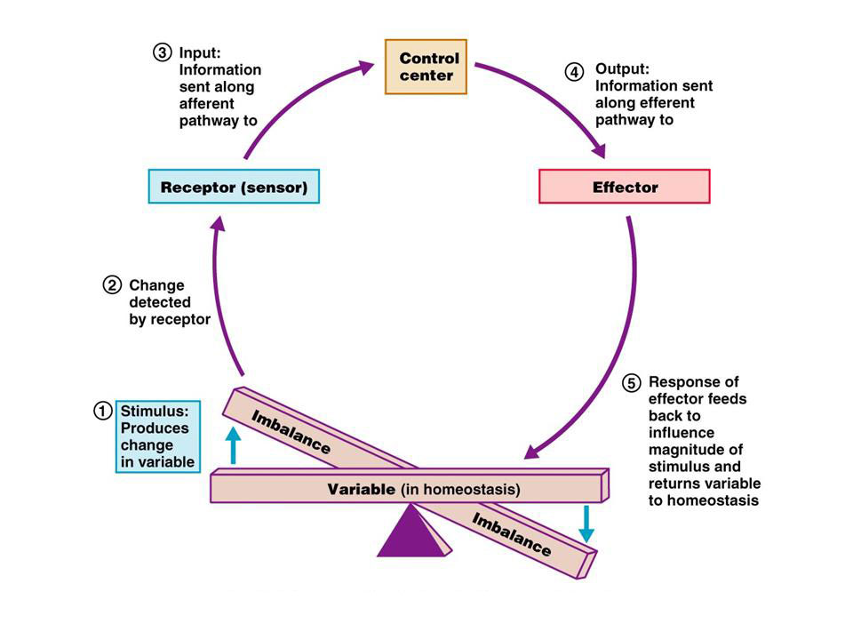
To maintain homeostasis, communication within the body is essential. The image below is an example of how a homeostatic control system works. Here is a brief explanation:
  1. Stimulus- produces a change to a variable (the factor being regulated).
  2. Receptor- detects the change. The receptor monitors the environment and responds to change (stimuli).
  3. Input- information travels along the (afferent) pathway to the control center. The control center determines the appropriate response and course of action.
  4. Output- information sent from the control center travels down the (efferent) pathway to the effector.
  5. Response- a response from the effector balances out the original stimulus to maintain homeostasis.
Image of a homeostatic control system using positive and negative feedback
Interactions among the elements of a homeostatic control system maintain stable internal conditions by using positive and negative feedback mechanisms.
Think of it as an extremely complex balancing act. Here’s a few more definitions you may want to know.
Afferent pathways- carry nerve impulses into the central nervous system. For instance, if you felt scorching heat on your hand, the message would travel through afferent pathways to your central nervous system.
Efferent pathways- carry nerve impulses away from the central nervous system to effectors (muscles, glands).
The feeling of heat would travel through an afferent pathway to the central nervous system. It would then interact with the effector and travel down the efferent pathway, eventually making the person remove their hand from the scorching heat.

Negative feedback mechanisms

Almost all homeostatic control mechanisms are negative feedback mechanisms. These mechanisms change the variable back to its original state or “ideal value”.
A good example of a negative feedback mechanism is a home thermostat (heating system). The thermostat contains the receptor (thermometer) and control center. If the heating system is set at 70 degrees Fahrenheit, the heat (effector) is turned on if the temperature drops below 70 degrees Fahrenheit. After the heater heats the house to 70 degrees Fahrenheit, it shuts off effectively maintaining the ideal temperature.
The control of blood sugar (glucose) by insulin is another good example of a negative feedback mechanism. When blood sugar rises, receptors in the body sense a change . In turn, the control center (pancreas) secretes insulin into the blood effectively lowering blood sugar levels. Once blood sugar levels reach homeostasis, the pancreas stops releasing insulin.
These are just two examples of negative feedback mechanisms within our body, there are 100’s, can you think of a few more?
Positive feedback mechanisms
A positive feedback mechanism is the exact opposite of a negative feedback mechanism. With negative feedback, the output reduces the original effect of the stimulus. In a positive feedback system, the output enhances the original stimulus. A good example of a positive feedback system is child birth. During labor, a hormone called oxytocin is released that intensifies and speeds up contractions. The increase in contractions causes more oxytocin to be released and the cycle goes on until the baby is born. The birth ends the release of oxytocin and ends the positive feedback mechanism.
Another good example of a positive feedback mechanism is blood clotting. Once a vessel is damaged, platelets start to cling to the injured site and release chemicals that attract more platelets. The platelets continue to pile up and release chemicals until a clot is formed.
Just remember that positive feedback mechanisms enhance the original stimulus and negative feedback mechanisms inhibit it.

Tuesday, 16 December 2014

Prion Diseases

A prion (Listeni/ˈpriːɒn/) is an infectious agent, specifically a protein in a misfolded form. The word prion, coined in 1982 by Stanley B. Prusiner, is derived from the words protein and infection. The protein itself, whether in its misfolded or its correctly folded form, can be referred to as the prion protein (PrP). A protein as an infectious agent stands in contrast to all other known infectious agents, like viruses, bacteria, fungi, or parasites—all of which must contain nucleic acids (either DNA, RNA, or both). Prions are responsible for mammalian transmissible spongiform encephalopathies, including bovine spongiform encephalopathy (BSE, also known as "mad cow disease") and scrapie in sheep. In humans, prions cause Creutzfeldt-Jakob Disease (CJD), variant Creutzfeldt-Jakob Disease (vCJD), Gerstmann–Sträussler–Scheinker syndrome, Fatal Familial Insomnia and kuru.[4] All known prion diseases in mammals affect the structure of the brain or other neural tissue and all are currently untreatable and universally fatal. In 2013, a study revealed that 1 in 2,000 people in the United Kingdom might harbour the infectious prion protein that causes vCJD.

Prions are not considered living organisms but are misfolded protein molecules which may propagate by transmitting a misfolded protein state. If a prion enters a healthy organism, it induces existing, properly folded proteins to convert into the disease-associated, misfolded prion form; the prion acts as a template to guide the misfolding of more proteins into prion form. These newly formed prions can then go on to convert more proteins themselves; this triggers a chain reaction that produces large amounts of the prion form. All known prions induce the formation of an amyloid fold, in which the protein polymerises into an aggregate consisting of tightly packed beta sheets. Amyloid aggregates are fibrils, growing at their ends, and replicating when breakage causes two growing ends to become four growing ends. The incubation period of prion diseases is determined by the exponential growth rate associated with prion replication, which is a balance between the linear growth and the breakage of aggregates. (Note that the propagation of the prion depends on the presence of normally folded protein in which the prion can induce misfolding; animals that do not express the normal form of the prion protein can neither develop nor transmit the disease.)

This altered structure is extremely stable and accumulates in infected tissue, causing tissue damage and cell death.[9] This structural stability means that prions are resistant to denaturation by chemical and physical agents, making disposal and containment of these particles difficult. Prions come in different strains, each with a slightly different structure, and, most of the time, strains breed true. Prion replication is nevertheless subject to occasional epimutation and then natural selection just like other forms of replication.

All known mammalian prion diseases are caused by the so-called prion protein, PrP. The endogenous, properly folded form is denoted PrPC (for Common or Cellular), whereas the disease-linked, misfolded form is denoted PrPSc (for Scrapie, after one of the diseases first linked to prions and neurodegeneration.)[11][12] The precise structure of the prion is not known, though they can be formed by combining PrPC, polyadenylic acid, and lipids in a Protein Misfolding Cyclic Amplification (PMCA) reaction. Proteins showing prion-type behavior are also found in some fungi, which has been useful in helping to understand mammalian prions. Fungal prions do not appear to cause disease in their hosts

Protein Structure and Protein Denaturation

Denaturation is a process in which proteins or nucleic acids lose the quaternary structure, tertiary structure and secondary structure which is present in their native state, by application of some external stress or compound such as a strong acid or base, a concentrated inorganic salt, an organic solvent (e.g., alcohol or chloroform), radiation or heat. If proteins in a living cell are denatured, this results in disruption of cell activity and possibly cell death. Denatured proteins can exhibit a wide range of characteristics, from conformational change and loss of solubility to communal aggregation.

This concept is unrelated to denatured alcohol, which is alcohol that has been mixed with additives to make it unsuitable for human consumption.

Reflex Arc


A reflex arc is a neural pathway that controls an action reflex. In higher animals, most sensory neurons do not pass directly into the brain, but synapse in the spinal cord. This characteristic allows reflex actions to occur relatively quickly by activating spinal motor neurons without the delay of routing signals through the brain, although the brain will receive sensory input while the reflex action occurs.

Reflexes in the Colon


The gastrocolic reflex or gastrocolic response is one of a number of physiological reflexes controlling the motility, or peristalsis, of the gastrointestinal tract. It involves an increase in motility of the colon in response to stretch in the stomach and byproducts of digestion in the small intestine. Thus, this reflex is responsible for the urge to defecate following a meal. The small intestine also shows a similar motility response. The gastrocolic reflex helps make room for more food.
The reflex was demonstrated by myoelectric recordings in the colons of animals and humans, which showed an increase in electrical activity within as little as 15 minutes after eating. The recordings also demonstrated that the gastrocolic reflex is uneven in its distribution throughout the colon. The sigmoid colon is more greatly affected than the right side of the colon in terms of a phasic response; however, the tonic response across the colon is uncertain. When pressure within the rectum becomes increased, the gastrocolic reflex acts as a stimulus for defecation. A number of neuropeptides have been proposed as mediators of the gastrocolic reflex. These include serotonin, neurotensin, cholecystokinin (CCK), and gastrin.

Clinically, the gastrocolic reflex has been implicated in pathogenesis of irritable bowel syndrome: the very act of eating or drinking can provoke an overreaction of the gastrocolic response in some patients with irritable bowel syndrome due to their heightened visceral sensitivity, and this can lead to abdominal pain, diarrhea, or constipation.[2] Also, the serotonin (5HT3) antagonist ondansetron decreases the tonic response to stretch.

Respiration


In physiology, respiration is defined as the transport of oxygen from the outside air to the cells within tissues, and the transport of carbon dioxide in the opposite direction.

The physiological definition of respiration should not be confused with the biochemical definition of respiration, which refers to cellular respiration: the metabolic process by which an organism obtains energy by reacting oxygen with glucose to give water, carbon dioxide and ATP (energy). Although physiologic respiration is necessary to sustain cellular respiration and thus life in animals, the processes are distinct: cellular respiration takes place in individual cells of the organism, while physiologic respiration concerns the bulk flow and transport of metabolites between the organism and the external environment.

Breathing (which in organisms with lungs is called ventilation and includes inhalation and exhalation) is a part of physiologic respiration. Thus, in precise usage, the words breathing and ventilation are hyponyms, not synonyms, of respiration; but this prescription is not consistently followed, even by most health care providers, because the term respiratory rate (RR) is a well-established term in health care, even though it would need to be consistently replaced with ventilation rate if the precise usage were to be followed.

Sarcomere Shortening


A sarcomere (Greek sarx "flesh", meros "part") is the basic unit of a muscle. Muscles are composed of tubular muscle cells (myocytes or myofibers), which are formed in a process known as myogenesis. Muscle cells are composed of tubular myofibrils. Myofibrils are composed of repeating sections of sarcomeres, which appear under the microscope as dark and light bands. Sarcomeres are composed of long, fibrous proteins that slide past each other when the muscles contract and relax.
Two of the important proteins are myosin, which forms the thick filament, and actin, which forms the thin filament. Myosin has a long, fibrous tail and a globular head, which binds to actin. The myosin head also binds to ATP, which is the source of energy for muscle movement. Myosin can only bind to actin when the binding sites on actin are exposed by calcium ions.

Actin molecules are bound to the Z line, which forms the borders of the sarcomere. Other bands appear when the sarcomere is relaxed.

A muscle cell from a biceps may contain 100,000 sarcomeres.The myofibrils of smooth muscle cells are not arranged into sarcomeres.

Monday, 15 December 2014

Sense of Balance Equilibrioception


Hearing


The sense of hearing is the ability to detect the mechanical vibrations we call sound.
Sound waves
  • pass down the auditory canal of the outer ear
  • strike the eardrum (tympanic membrane) causing it to vibrate
  • these vibrations are transmitted across the middle ear by three tiny linked bones, the ossicles:
    • hammer (malleus)
    • anvil (incus)
    • stirrup (stapes)
  • The ossicles also magnify the amplitude of the vibrations.
The middle ear is filled with air and is connected to the outside air by the eustachian tube, which opens into the nasopharynx. Opening of the tube — during swallowing or yawning — equalizes the air pressure on either side of the eardrum.
Allergies or a head cold may inflame the walls of the eustachian tubes making them less easily opened. Rapid changes in pressure at such times — such as descending in an aircraft or during a SCUBA dive, may be quite painful because of the unequal pressure against the eardrums.

The Inner Ear

Vibrations of the innermost ossicle, the stirrup, are transmitted through a flexible membrane, the oval window to the cochlea of the inner ear.
The cochlea is a tube, about 3.5 cm long, that is coiled like a snail shell and filled with a special fluid called endolymph. The most dramatic difference in the composition of endolymph from other lymph in the body is its high concentration of potassium (K+) ions.
Running through the cochlea for its entire length is a plate of bone and an inner tube that is also filled with endolymph. These structures divide the outer tube of the cochlea into two separate chambers.
Because liquids are practically incompressible, it is necessary to have some way of relieving the pressures created when the oval window is pushed in and out. The flexible round window does this by moving in the opposite direction.

The organ of Corti

The organ of Corti lies within the middle chamber of the cochlea. It contains thousands of hair cells, which are the actual vibration receptors. The apical surface of the hair cells contains an array of stereocilia, which give the hair cells their name. Stereocilia are not built from the "9+2" arrangement of microtubules that are found in true cilia.
The hair cells are located between the basilar and tectorial membranes. Vibrations of the endolymph cause vibrations of the basilar membrane. This moves stereocilia at the tips of the hair cells against the tectorial membrane and open potassium channels in them. The influx of K+ from the endolymph depolarizes the cell.
You should note that hair cells differ from most "excitable cells" (neurons and muscle fibers) in their use of potassium ions, not sodium ions, to depolarize the cell.
Link to a discussion of excitable cells.
Depolarization of the hair cell causes the release of a neurotransmitter (probably glutamate) at its basal surface and the initiation of nerve impulses in a sensory neuron that synapses with it. These impulses travel back along the auditory nerve (the 8th cranial nerve) to the brain.
Many people, especially when young, can hear sounds with frequencies (pitches) from as low as 16 to as high as 20,000 hertz (cycles per second). Detection of a given frequency is a function of the location of the hair cells along the organ of Corti with the highest frequencies detected near the base of the cochlea, and the remainder of the sound spectrum detected in a progressive fashion with the lowest frequencies detected by hair cells near the tip.

Deafness

Deafness may be acquired or inherited.

Acquired deafness

If a laboratory animal is exposed to very intense, pure tones, it eventually becomes deaf to those frequencies, but its ability to hear other pitches is unimpaired. Examination of its organ of Corti reveals destroyed hair cells in a single area whose location can be easily correlated with the pitch of the destructive sound.
Similar deficits occur in humans who are exposed to intense noises for long periods. (A trained audiologist can tell by looking at the frequency response whether a patient flies private aircraft.)

Inherited deafness

About 1 newborn in a thousand is born deaf because of a genetic defect. As the years go by, many of us (~16%) suffer a progressive loss of hearing because of genetic defects.
Literally scores of genes have been identified in recent years whose mutant versions result in hearing loss.
  • mutations in a transcription factor have been associated with a stirrup (stapes) that cannot move freely and thus cannot transmit vibrations to the oval window.
  • The proper organization of the stereocilia involves actin, a form of myosin (called myosin VIIA), and cadherins.
    • Mutations in the gene encoding a protein that helps with actin polymerization cause deafness.
    • Mutations in the myosin VIIA gene and
    • mutations in the gene encoding cadherin 23
    also cause deafness.
  • The potassium that enters the hair cells must be removed from them and recycled back to the endolymph for hearing to continue. Scores of mutations in the necessary transport molecules have been linked to inherited deafness.
    • Mutations in genes encoding the K+ channels that allows K+ to leave the hair cell through its basolateral surface (shown in green ). (These same channels are found in the loops of Henle in the kidneys so the mutations can produce defects in kidney function as well as deafness.)
    • Mutations in the connexins (magenta) that form the gap junctions through which the K+ passes from cell to cell on its way back to the secretory cells that will deposit it back in the endolymph.
      Link to a discussion of gap junctions and connexins.
    • Mutations in the sodium-potassium-chloride cotransporter (shown in yellow) that actively transports K+ against its concentration gradient into the secretory cells.
      Link to discussion of active transport.
    • mutations in the K+ channels (shown in lavender) that allow for the facilitated diffusion of K+ out of the secretory cells and into the endolymph.

Equilibrium

The inner ear also detects:
  • the position of the body with respect to gravity
  • the motion of the body.
Just above the cochlea are two interconnecting chambers filled with endolymph, the sacculus and utriculus. On their inner surface are patches of hair cells to which are attached thousands of tiny spheres of calcium carbonate (CaCO3). Gravity pulls these downward. As the head is oriented in different directions, these ear stones or otoliths shift their position. The action potentials initiated in the hair cells are sent back to the brain.
Motion of the body is detected in the three semicircular canals at the top of each inner ear, each one oriented in a different plane. There is a small chamber at one end of each canal containing hair cells. Whenever the head is moved, the fluid within the canals lags in its motion so that there is relative motion between the walls and the endolymph. This stimulates the hair cells to send impulses back to the brain.
When the hair cells send messages that are incongruent with what the eyes are seeing and our body is feeling, as may occur in a boat or aircraft during rough weather, motion sickness can result.
Some people also suffer severe dizziness because otoliths have become dislodged from their utriculus (e.g. following a blow to the head) and settled in a semicircular canal.

Echolocation

Bats can hear frequencies as high as 150,000 hertz. Sound at these ultrasonic (to us) frequencies travels in fairly straight lines. Bats flying in complete darkness are able to locate obstacles and even insect prey by emitting pulses of this ultrasonic sound and then adjusting the course of flight to the echo that returns to their ears. Such a system of echolocation works on the same principle as sonar for submarine detection.
A blindfolded bat can fly between the wires touching them only rarely. A bat whose ears are plugged collides repeatedly with the wires.
Hunter and hunted. In the top photo, a moth (bright streak) takes successful evasive action upon detecting the approach of a bat (broad streak across the photo). (The diffuse image is a tree in the background.) In the bottom photo, the two streaks intersect, indicating that this time the moth was unable to escape capture by the bat. (Photos by Frederic A. Webster, courtesy of the late Prof. Kenneth D. Roeder.)